This is an automatically translated article.
The article is professionally consulted by Master, Doctor Cao Thanh Tam - Cardiologist - Cardiovascular Center - Vinmec Central Park International General Hospital.Echocardiography is a non-invasive, safe and widely used imaging method in the identification of cardiac abnormalities.
1. What is echocardiography?
Echocardiography is a noninvasive exploratory technique that uses ultrasound waves to identify abnormalities in the structure and function of the heart.
Ultrasound waves are emitted during the procedure, it travels in and out of your heart and creates a moving image of it on the screen. This allows your doctor to look at your heart's anatomy from many different angles and monitor your heart rate.
Based on the principle of operation, echocardiography is divided into 3 types:
1 dimensional ultrasound: A form of exploration of the anatomical components of the heart; 2-D Ultrasound: Aims to clearly see the anatomical sandstones of the heart and the images seen will be almost like the real anatomy; Doppler ultrasound: Helps to survey the changes in morphology, function as well as hemodynamics of the heart. Based on location, there are 2 types of ultrasound:
Transthoracic echocardiography: This is the most common echocardiographic technique. The probe is placed on the outside of the chest wall. Then, the doctor will apply gel to the chest to help the sound waves travel better. The transducer uses ultrasound waves through the chest and creates an image of the heart on a connected computer monitor. Transesophageal echocardiography: This method is rarely used, the doctor will use a thinner probe attached to the end of the endoscope. This tube will be inserted into the esophagus to help the doctor better see the details of the heart from behind.

Siêu âm tim qua thành ngực là kỹ thuật phổ biến nhất
Echocardiography technology plays an important role in screening and diagnosing the patient's cardiovascular health, helping:
Provide accurate information about the shape of the heart such as the size of the heart chambers , condition of the heart valves, wall thickness of the heart... Provides information about left atrial, right atrial, left ventricular, right ventricular function (in which, the role of assessing left ventricular function is important. and has the most applications in the screening and treatment of cardiovascular diseases).
2. Basic views in thoracic echocardiography
2.1 Subcostal view The transducer is perpendicular to the abdominal wall at the position below the sternum blade, pointing to the left. The subcostal view gives orientation to the arrangement of intra-abdominal viscera, helping to evaluate the patient's 4 chambers of the heart.
2.2 Parasternal view Carrying out exploration of the position of the parasternal view helps to evaluate the left ventricle, compare the size of the heart chambers and localize pericardial effusion, distinguish pericardial effusion and pleural effusion.
2.3 Short-axis sternal view Probe the short-axis sternal position to obtain images by rotating the transducer 90 degrees clockwise, then scanning the transducer from the base of the heart up to the apex to obtain different cross-sections.

Có 4 mặt cắt cơ bản trong siêu âm tim thành ngực
2.4 Apical view Performing apical exploration is considered to be more difficult, but this technique is effective in comparing ventricular chamber sizes and is the best window for assessing motor abnormalities. in the interventricular septum and the heart wall.
3. Things to know about echocardiography
Before the ultrasound, you can still eat and drink completely normal. However, if you need a transesophageal or stress ultrasound, your doctor will ask you to fast for several hours before the ultrasound. You will lie on the bed and pull your shirt up. Then, your doctor will apply a special gel on your chest to increase the conduction of the sound waves and remove air between the skin and the transducer (the transducer is a small plastic device that sends and receive sound waves). The doctor moves the transducer back and forth across the chest, and the sound waves that create an image of the heart are recorded on a computer screen. If you're having a transesophageal echocardiogram, you'll be numbed with an inhaler or sedated to make it easier for the transducer to get into your esophagus. Then, the endoscope is inserted into your throat. The doctor will push the fistula deep into the esophagus and take pictures of the right and left chambers of the heart.
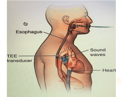
Hình ảnh siêu âm tim qua thực quản
So far, there have been no recorded side effects of echocardiography, therefore, this is still a safe and commonly used imaging method, echocardiography is a non-invasive technique. Therefore, the patient does not feel any pain during the procedure.
Currently, there are many medical facilities that apply ultrasound technology in the early diagnosis and treatment of diseases. Vinmec International General Hospital system has been using the most modern generations of color ultrasound machines today in examining and treating patients.
One of them is GE Healthcarecar's Logig E9 ultrasound machine with full options, HD resolution probes for clear images, accurate assessment of lesions.
Besides, a team of experienced doctors and nurses will greatly assist in the diagnosis and early detection of abnormal signs of the body in order to provide timely treatment.
To register to perform echocardiography at Vinmec International General Hospital, please book an appointment on the website for service.
Please dial HOTLINE for more information or register for an appointment HERE. Download MyVinmec app to make appointments faster and to manage your bookings easily.