This is an automatically translated article.
The article was professionally consulted by Specialist Doctor I Vo Cong Hien - Radiologist - Radiology Department - Vinmec Nha Trang International General Hospital and Master, Doctor Le Xuan Thiep - Doctor Doctor of Radiology - Department of Diagnostic Imaging - Vinmec Ha Long International General Hospital.Percutaneous drainage of the renal pelvis under the guidance of digital background erasure imaging helps to quickly resolve the stagnant water and pus in the patient's renal pelvis. This procedure is used when the urinary tract is blocked for various reasons.
1. What is percutaneous pyelonephritis?
Percutaneous pyelonephritis is a method of establishing a path to drain urine from the renal pelvis to the outside of the skin in order to resolve water stagnation and pus in the renal pelvis. Percutaneous pyelonephritis is indicated in case the upper urinary tract is blocked due to causes such as:Urinary stones, pyelonephritis - ureteral stricture after surgery, pyelonephritis pyelonephritis. , sclerosing ureteritis . Malignant diseases causing obstruction of the urinary tract such as uterine cancer, urinary system cancer, prostate cancer, metastatic pelvic cancer. Obstruction of the urinary tract during pregnancy, the cause has not been completely resolved. Under the guidance of imaging methods, a specialized drainage tube will be inserted into the renal pelvis to lead urine out. Percutaneous pyelonephritis is a method with high safety and immediate effect, helping to resolve local infections, limiting the risk of life-threatening infections spreading and at the same time for other patients. The doctor can give the doctor more time to deal with the cause of the blockage.
However, there are also some cases where it is not possible to immediately perform percutaneous pyelonephritis. That is when patients have severe coagulopathy, severe electrolyte disturbances, hypertension,... these patients need to be stabilized before intervention.
Besides, in some cases it is necessary to promptly examine and treat the following cases.
Urinary stones, pyelonephritis - ureteral stricture after surgery, pyelonephritis pyelonephritis, sclerosing ureteritis. Malignant diseases causing obstruction of the urinary tract such as uterine cancer, urinary system cancer, prostate cancer, metastatic pelvic cancer. Obstruction of the excretory tract during pregnancy, the cause has not been completely managed, prioritizing percutaneous pyelonephritis drainage under ultrasound guidance.
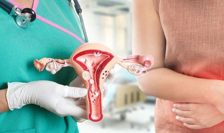
Bệnh nhân ung thư tử cung được chỉ định thực hiện thủ thuật
2. Application of digital background erasure imaging in percutaneous pyelonephritis
Percutaneous pyelonephritis must be performed under the guidance of imaging methods. The media that can be used are ultrasound and fluorescent screens or digital background removal imaging (DSA). In particular, DSA is a modern tool that can be used in many stages of the percutaneous pyelonephritis drainage process such as: helping to determine the location to put the drainage into the renal pelvis, guiding the direction of the renal pelvis. guide during the insertion of the catheter and check that the position of the catheter in the renal pelvis is correct.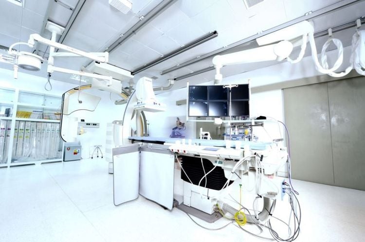
Chụp số hóa xóa nền DSA giúp chẩn đoán hình ảnh cho dẫn lưu bể thận qua da
3. Percutaneous pyelonephritis drainage under the guidance of digital background erasure
3.1. Prepare
The implementation team includes specialist doctors, assistant doctors, radiology technicians and nurses. In case the patient cannot cooperate, additional support from the doctor or anesthesiologist is needed.The necessary facilities are a digital background eraser (DSA); film, film printer, image storage system; lead vest, apron, X-ray shielding; anesthetics, anesthetics, and contrast agents. In addition to common medical supplies such as syringes, cotton, bandages, gauze, ... also need special medical supplies such as chiba needles, vascular catheter sets, rigid leads, angiographic catheters, etc. ..
On the patient's side, the medical staff will advise on the current health situation, explain the purpose, effects and complications of the procedure. Patients must fast before the procedure for at least 6 hours, can drink water but not more than 50ml.
3.2. Steps to take
The team installed a machine to monitor blood pressure, breathing rate, electrocardiogram, SpO2 for the patient. The patient is tested for response to the anesthetic. The patient lies on his side to expose the kidney to be drained. The doctor washes his hands, wears sterile gloves, disinfects the area to be drained, and then covers with a sterile cloth with holes. The doctor conducts anesthesia in layers, makes incisions in the skin and subcutaneous tissue into the lumbar fossa behind the peritoneum. Under the guidance of digital subtraction angiography (DSA), percutaneous pyelonephritis was performed with a 21G needle. Check the position of the needle tip in the renal pelvis with contrast. Create a tunnel into the renal pelvis through the skin by: inserting a rigid 0.018 wire into the renal pelvis through the 21G needle, then replace the 21G needle with a tube into the lumen. Replace the 0.018 wire with 0.035 J-tipped wire, then insert the catheter into the renal pelvis. Perform pyelonephritis imaging downstream through the catheter. Use the tubes to tunnel through the 0.035 J-tipped wire. Begin catheterization by inserting a pigtail into the renal pelvis through 0.035 J-tipped wire. Check the position of the drainage tube tip with contrast through the image of the digital background eraser. After determining that the tip of the drain is in the correct position, the drain is fixed in the renal pelvis and outside the skin. The procedure of percutaneous pyelonephritis drainage under the guidance of digital erasure is evaluated as successful when the tip of the drain is placed in the renal pelvis, the pyelonephritis is collapsed and there is urine out through the drain. Contrast does not escape into the abdominal cavity nor into the retroperitoneal space.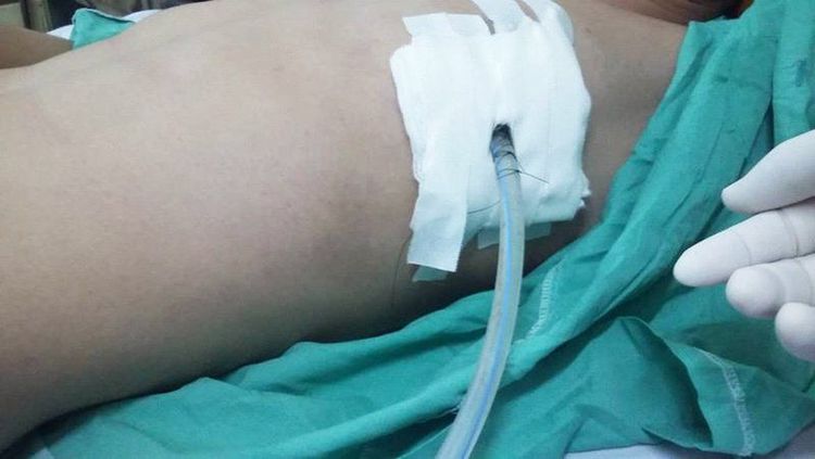
Đầu ống dẫn lưu bằng thuốc đối quang qua hình ảnh của chụp số hóa xóa nền cần được kiểm tra vị trí
4. Possible complications when percutaneous pyelonephritis drainage under the guidance of digital background removal
Although a safe procedure, infection can occur with percutaneous pyelonephritis. There are degrees of infection, which can be local, urinary tract infection, or bacteremia. If there is an infection at the drainage site, treat it by cleaning, changing the dressing, and disinfecting the infected site. If a urinary tract infection occurs, the patient will be treated with systemic antibiotics. Sepsis is a dangerous, life-threatening condition, a specialist consultation will be performed to find a treatment plan for the patient.Another possible complication is pyelonephritis - ureteral bleeding, medical staff will manually squeeze at the lumbar hole with drainage for 15-20 minutes. If bleeding continues, transfer to the intervention room to replace with a larger size drain. If bleeding persists after drain replacement, vascular intervention is performed.
Percutaneous pyelonephritis is a technique that requires a high level of experience of the doctor and the perfect coordination of the patient. In addition, in order to achieve high diagnostic efficiency, patients need to choose reputable addresses with digital scanners to erase the background, modern and standard medical equipment.
Vinmec International General Hospital is a hospital with a full convergence of general and specialized doctors to perform, examine, operate, diagnose and treat kidney and urinary diseases. In particular, at Vinmec, percutaneous pyelonephritis is also performed to treat many different diseases and provide optimal treatment results for customers.
BSCK I Vo Cong Hien has many years of experience in the field of imaging, especially in diagnostic ultrasound for general abdominal diseases, advanced cardiac - vascular, obstetrics and gynecology. Doctor Vo Cong Hien used to work at the Imaging Department of the University of Medicine and Pharmacy Hospital - Hoang Anh Gia Lai before working at the Imaging Department of Vinmec Nha Trang International General Hospital.
Dr. Le Xuan Thiep has strengths in performing advanced and difficult magnetic resonance and computed tomography techniques such as: coronary computed tomography, cardiac function, cerebral magnetic resonance, perfusion brain and organs,..
If you notice any unusual health problems, you should visit and consult with a specialist.
Please dial HOTLINE for more information or register for an appointment HERE. Download MyVinmec app to make appointments faster and to manage your bookings easily.


