This is an automatically translated article.
Article by Dr. Vu Thi Hanh, Doctor of Radiology - Department of Diagnostic Imaging - Vinmec Hai Phong International General Hospital.Depending on the time of pregnancy, fetal ultrasound may be indicated for different purposes. Fetal enlargement of the bladder is known as an enlarged bladder and ureter observed on ultrasound.
1. Purpose of fetal ultrasound
During the first 3 months, fetal ultrasound is done to:
Confirm the mother's pregnancy, determine the gestational age and estimate the date of delivery Check the fetal heart rate, detect abnormalities if any Check the Possible abnormalities of placenta, uterus, ovaries... Diagnosis of ectopic pregnancy or risk of miscarriage During the second and third trimesters of pregnancy :
Monitor growth and location of the fetus Determine the sex of the baby Check the placenta for placental covering or placental abruption

Thai ngoài tử cung
Check for features of Down syndrome (usually done between 12-13 weeks 6 days of gestation) Check for birth defects or abnormalities in the structure or blood flow of the fetus, identify the fetus getting enough oxygen Diagnose problems with the mother's ovaries or uterus Measure cervical length or instruct other techniques such as amniocentesis Confirm fetal death
2. Based on what factors to diagnose fetal bladder enlargement – Megacystis
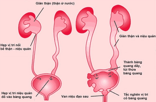
Fetal enlargement is known as: enlarged bladder and ureters observed on ultrasound. Prenatal detection rate is 1 in 1500 pregnant women, more often boys than girls are detected (ratio 8/1). During the first trimester of pregnancy, cystitis is diagnosed when the bladder length is greater than 7mm.
Normal bladder height < 7mm; When the bladder height is from 7-15mm: 20% aneuploidy; 10% urinary tract pathology; 70% normal. When bladder height > 15mm: 10% aneuploidy; 90% of urinary tract diseases.
3. Causes of an enlarged bladder in the fetus

Van niên đạo
Posterior urethral valve (57%). Urethral stricture and atrophy (7%) Prune belly syndrome (4%). Magacystis – microcolon- untestinal-hypopestalsis syndrome (MMIHS) (1%). Abnormalities of the disk (0.7%). Chromosomal abnormalities (15%) include chromosomes 21, 18, and 13.
4. What should pregnant women do when the fetal bladder is diagnosed on ultrasound?
Pregnant women need to be monitored to have the most appropriate management attitude according to the advice of a specialist. of kidney, karyotype and sex About 50% of pregnancy termination Vinmec International General Hospital offers a Package Maternity Care Program for pregnant women from the very first months of pregnancy with full regular antenatal check-ups, 3D and 4D ultrasounds and routine tests to ensure that the mother is healthy and the fetus develops comprehensively. Pregnant women will no longer be alone when entering labor because having a loved one to help them during childbirth always brings peace of mind and happiness.
Please dial HOTLINE for more information or register for an appointment HERE. Download MyVinmec app to make appointments faster and to manage your bookings easily.


