This is an automatically translated article.
The article was professionally consulted by Specialist Doctor I Le Hong Lien - Department of Obstetrics and Gynecology - Vinmec Central Park International General Hospital.The 20th week ultrasound is an important transition period in your pregnancy, so it is very important to understand the physical and physiological changes of mother and baby and what to prepare for
1. What will we see in the ultrasound?
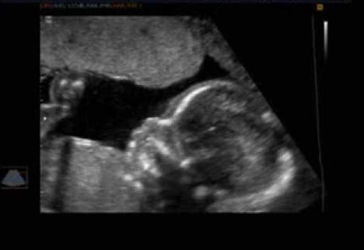
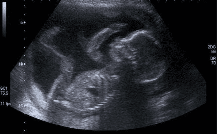
With a 20-week ultrasound you can get a better idea of what your baby will be like. But there's more to it than that. At this point in pregnancy, although the fetal organs are still immature, all of them are formed, including all of the heart's outflows and outflows - the vena cava, the aorta, and the aorta. pulmonary artery.The purpose of a 20 to 22 week ultrasound is to review all of the fetal anatomy and to determine if all is normal. Things that can't be seen on those ultrasounds, such as spinal cord abnormalities, brain defects, heart defects, and diaphragmatic abnormalities, can often be seen on this ultrasound.
This is also the time to measure the baby's growth, to make sure that nothing is wrong. The size of the fetus measured during the ultrasound will fall within the expected range for gestational age. A doctor may order additional testing if your child's growth is out of the predicted range. And if a problem is identified, specific recommendations can be made for prospective parents, who can then meet with the appropriate specialists and the birth can be planned at a hospital.
A 20-week ultrasound can also be helpful in ensuring a healthy and fruitless delivery for you. Details of the uterus, placenta and amniotic fluid can be seen. If your doctor notices anything unusual, he can make recommendations regarding your birth plan to help ensure that you and your baby are safe.

2. What can pregnant women see on ultrasound?
Most hospitals allow you to see during the ultrasound. If you haven't had an ultrasound during your pregnancy, the sonographer will check.The sonographer will show the baby's heart rate and body parts, such as the face and limbs, toes and feet, the movements of the feet and hands, before looking at the baby in detail. Your baby's bones will appear white when scanned and his soft tissues will be gray and mottled. The amniotic fluid surrounding your baby will look black.
After you see your baby on the screen, some sonographers will rotate the screen away for the rest of the ultrasound and show you the view at the end.
3. Ultrasound parameters of pregnant women
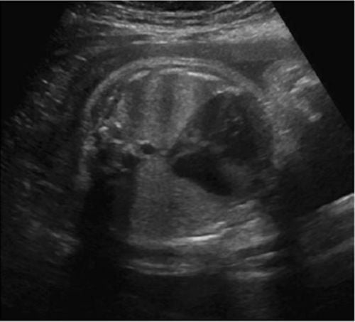
Ultrasound doctor gives pregnant woman 20 weeks pregnant to check all the baby's organs for abnormalities and take measurements. The doctor will read the results:Shape and structure of your baby's head and brain. At this stage, serious brain problems are rare. Baby's face, to check for cleft lip. Cleft palate inside a baby's mouth is difficult to see and is not usually visible. The baby's spine both along its length, and in cross section, to ensure that all the bones are in alignment, and that the skin covers the spine in the back. Your baby's abdominal wall, to make sure it covers all the internal organs in the front. In a baby's heart, the top two chambers (atria) and the bottom two chambers (ventricles) should be of equal size. The valves should open and close with each heartbeat. The sonographer will also examine the major veins and arteries that carry blood to and from your baby's heart. Baby's stomach. The baby swallows some of the amniotic fluid on which he lies, seen in his abdomen as a black bubble.

4. Abnormal readings can be seen on ultrasound
A 20-week fetal ultrasound will look at your baby's bones, heart, brain, spinal cord, face, kidneys, and abdomen in detail. It allows the sonographer to look for 11 rare conditions. The ultrasound only looks for these conditions and can't find everything that could go wrong.You can find more information about each condition, including treatment options: asthenia, open spine, cleft lip, diaphragmatic hernia, digestive, exomphalos, serious heart abnormalities, spasms bilateral renal failure, fatal skeletal dysplasia, Edwards Syndrome, or T18, Patau Syndrome, or T13.
In most cases, the scan will show that the baby seems to be developing as expected, but sometimes the sonographer will find or suspect something different.

5. What if there are abnormal signs on the ultrasound?
Most problems that require repeat ultrasounds are not serious. About 15% of ultrasounds will be repeated for one reason or another. This could be because your baby is not in a good position, or you are overweight, in which case the ultrasound should be repeated at 23 weeks.If your sonographer finds or suspects a problem, you will be notified immediately. You should have an appointment for an ultrasound with a fetal medicine specialist within three to five days.
If the specialist thinks your baby has a heart problem, he or she will ask you to come in for an echocardiogram. A fetal echo scan will take a detailed look at your baby's heart.
If any ultrasound reveals a serious problem, you will be greatly assisted to guide you through all the options. Although serious problems are rare, some families face the most difficult decision of all, whether to continue with the pregnancy.
Other problems could mean your baby needs surgery or treatment after birth, or even surgery while he's still in your womb. There will be a range of people supporting you through this, obstetricians, pediatricians, physical therapists.
6. Can ultrasound harm the fetus?
There's no risk to the baby or you with an ultrasound, but it's important to think carefully about whether to have an ultrasound.It can provide information that means you have to make some important decisions. For example, you may be offered follow-up tests that carry a risk of miscarriage and you will need to decide whether to have these tests done.
Vinmec International General Hospital offers a Package Maternity Care Program for pregnant women right from the first months of pregnancy with a full range of antenatal care visits, periodical 3D and 4D ultrasounds and routine tests to ensure that the mother is healthy and the fetus is developing comprehensively. Pregnant women will no longer be alone when entering labor because having a loved one to help them during childbirth always brings peace of mind and happiness.
Specialist I Le Hong Lien is an obstetrician-gynecologist at Vinmec Central Park International Hospital since November 2016. Doctor Lien has over 10 years of experience as a radiologist in the Department of Ultrasound at the leading hospital in the field of obstetrics and gynecology in the South - Tu Du Hospital.

Please dial HOTLINE for more information or register for an appointment HERE. Download MyVinmec app to make appointments faster and to manage your bookings easily.
Articles refer to sources: parents.com, babycentre.co.uk, nhs.uk


