This is an automatically translated article.
The article was professionally consulted by Specialist Doctor I Tran Cong Trinh - Radiologist - Radiology Department - Vinmec Central Park International General Hospital.
Vascular ultrasound is a widely used and trusted method in the diagnosis of many diseases. So what exactly is the meaning of vascular ultrasound?
1. What is Vascular Ultrasound?
Vascular ultrasound is a common method applied to examine and evaluate the circulatory system in the body, locate the blockage caused by blood clots. Vascular ultrasound is a safe test that does not use ionizing radiation, providing detailed images of soft tissues that cannot be shown on an X-ray. Therefore, vascular ultrasound does not have any adverse effects on the patient's health.Vascular ultrasound simulates the activity of arteries and veins through images on the screen. When we do ultrasound, we can observe the structure and size of arteries and veins as well as investigate whether the blood flow inside is abnormal or not?
2. Meaning of vascular ultrasound
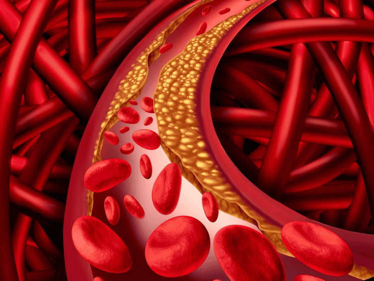
Vascular ultrasound is one of the techniques to help doctors check the flow of blood in the body. From there, it is possible to identify abnormalities such as arterial and venous obstruction, atherosclerosis, blood clots... fit.
Siêu âm mạch máu giúp kiểm tra những bất thường trong dòng chảy của máu
Specifically, vascular ultrasound is indicated to:
Monitor the flow of blood to organs and tissues in the body. Helps to detect the location of blockages and abnormal plaques and then supports the patient's treatment plan. Detect the location of blood clots. Assess whether the patient is able to undergo procedures such as angioplasty? Evaluation of graft or vascular bypass surgery. Determine if patient has vascular problems such as abdominal aortic aneurysm? Determine the origin and severity of varicose veins.
3. Ultrasound of blood vessels by new Doppler method
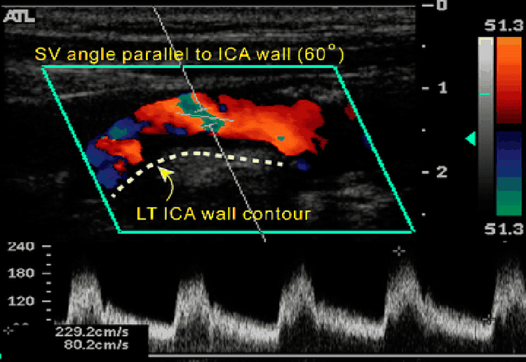
Doppler ultrasound is also a form of vascular ultrasound. This method is being applied by many people because it gives more accurate results than conventional ultrasound.Doppler ultrasound has the ability to survey the movement of objects with a transducer that receives ultrasound waves. Through the signals from the machine's transducer and the received frequency when surveying moving objects, the machine will synthesize the results on the screen in the form of colors, different running waveforms or audio signals. characteristic can be heard.
Doppler vascular ultrasound is a safe, completely painless test method, which is a tool to help doctors easily observe, monitor and evaluate the blood circulation system through arteries and veins. main circuit in the human body.

4. What cases need vascular ultrasound?
Patients are medically applied vascular ultrasound method when there is suspicion of diseases such as:Deep vein thrombosis: This is a disease related to blood clots in the veins. located deep inside the body, this is most often the case in the veins in the legs. Arteriosclerosis: The arteries that supply blood to the legs and feet harden and narrow, affecting blood flow to the lower body. Superficial thrombophlebitis: caused by blood clots forming in superficial veins, located just below the surface of the skin. Thromboangiitis is a condition in which the blood vessels of the hands and feet become inflamed and swollen. Vascular tumor located in the arm or leg.
5. What to prepare before vascular ultrasound?
In order to perform vascular ultrasound as quickly as possible, saving time, before you go you need to prepare:Wear loose-fitting clothes so that you can take them off as soon as the ultrasound doctor asks. Do not wear a lot of jewelry because jewelry can make you entangled during the ultrasound. Lying down on the hospital bed, restricting movement during the ultrasound.

Tháo bớt trang sức trước khi tiến hành siêu âm
6. Procedure for performing vascular ultrasound
The procedure to perform vascular ultrasound includes the following steps:First, the patient will be assigned by the doctor to lie on his side or face down on the examination table. Next is to apply water gel to the areas where the ultrasound needs to be performed to help the transducer make safe contact with the body. This step eliminates air pockets between the transducer and the skin that could block the sound waves from entering the body. Move the probe over the surface of the skin and at the same time the doctor reads the results on the projection screen. After the ultrasound is done, the patient changes clothes and waits for the doctor to review the ultrasound image. An ultrasound usually takes about 15-30 minutes. However, depending on the case, this process can take longer.
Conclusion: Currently, diseases related to circulation and veins are becoming more and more complicated, so vascular ultrasound becomes more and more important. As soon as you notice abnormal symptoms, go to a reputable hospital for examination and ultrasound to find out timely treatment.
Vinmec International General Hospital is one of the hospitals that not only ensures professional quality with a team of leading medical doctors, modern equipment and technology, but also stands out for its examination and consultation services. comprehensive and professional medical consultation and treatment; civilized, polite, safe and sterile medical examination and treatment space.
Before taking a job at Vinmec Central Park International General Hospital, the position of Doctor of Radiology from September 2017, Doctor Tran Cong Trinh worked at Gia Dinh People's Hospital since 2007. -2017. In his role, Dr. Tran Cong Trinh has participated in guiding the teaching of students, residents, specialists and new doctors entering the department
For examination and treatment at International General Hospital Vinmec, please come directly to Vinmec Health System or register online HERE.


