This is an automatically translated article.
The article is professionally consulted by Master, Doctor Nguyen Van Phan - Head of Interventional Imaging Unit - Department of Diagnostic Imaging and Nuclear Medicine - Vinmec Times City International General Hospital.There are many methods to examine cerebral blood vessels such as digital cerebral angiography with DSA background deletion, cerebral magnetic resonance angiography with and without contrast injection, computed tomography cerebral angiography with contrast injection. . Computed tomography cerebral angiography with contrast injection is a non-invasive method of high value in accurately diagnosing cerebrovascular diseases and brain parenchymal diseases.
1. Meaning of cerebral vascular computed tomography?
Brain vascular computed tomography is using a system of multi-detector or multi-layer computed tomography (64 layers, 128 layers, 256 layers, 512 cuts...) with contrast injection. Intravenous iodized radiograph to scan the entire cerebrovascular system and then use software to erase bone, software and reconstruct 3D images of cerebral blood vessels.From the images taken, it will assist the doctor in accurately diagnosing some diseases such as cerebral aneurysm, cerebral vascular malformation, cerebral stenosis or cerebral arteriovenous catheterization. From there, the doctor will give advice to the patient about the risk of accidents and complications caused by diseases such as: ruptured cerebral blood vessels, cerebral embolism (also known as stroke)...
In the emergency, when the patient comes to the hospital with a diagnosis of early cerebral stroke (before 6 hours from the onset of symptoms), the patient will be immediately assigned a CT scan of the brain with contrast injection. find the point of cerebral embolism due to blood clot and administer thrombolytic injection and intervene to remove the cerebral artery mechanical thrombus immediately. If the patient is determined to be bleeding in the brain, a computed tomography (CT) scan of the brain with contrast injection will be indicated to find the point of brain rupture (cerebral hemorrhage is usually due to the rupture of an aneurysm) and conduct internal intervention. embolized cerebral aneurysm by metal coils (coils). If the patient is diagnosed with other diseases without intervention, the patient is admitted to the hospital for treatment with a regimen appropriate to the patient's condition.

Phương pháp chụp cắt lớp vi tính mạch máu não giúp chẩn đoán chính xác một số bệnh lý
2. What are the risks of CT angiography of the brain?
If the patient is in an emergency situation of stroke or brain hemorrhage, the patient will be given a CT scan of the brain with contrast injection as soon as possible and closely monitored by emergency doctors. If the patient arrives in a non-emergency state of stroke, the safety regulations for injecting iodine-containing contrast agents should be observed as follows:Patient or guardian provide complete and accurate information the information in the safety checklist for injecting contrast for medical staff and signing the consent form for contrast injection. The information to be provided is as follows:
○ Have a history of allergies to weather, food, drug allergies, especially allergies to the ingredients of contrast in the previous scan.
○ Have kidney failure, severe heart failure.
○ Do you have asthma and are using your medication more than 2 times a day?
○ Are you experiencing severe dehydration?
○ The patient is diabetic and is being treated with the diabetes drug Metformin.
○ Have you fasted for 4 hours before?
○ For women of childbearing age, it is necessary to exploit information about whether they are pregnant, suspected of being pregnant or breastfeeding.
● The patient will be instructed to change into the computerized tomography room. Remove all jewelry, metal objects in the neck, head area so as not to affect the image quality and the shooting process.
● If the patient is using sedatives, they must have someone to support them and use appropriate means of transportation.
3. What to prepare before conducting CT angiography?
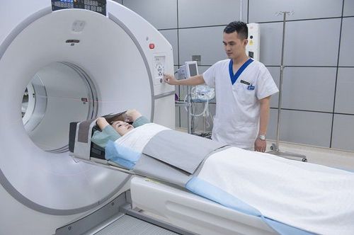
When taking pictures, the technician will explain the procedure of taking and injecting drugs for the patient to coordinate. The technician places an injection into a vein in the patient's elbow area and will connect it to a previously prepared system of injecting contrast agents containing iodine. The technician will put a light bandage on the patient's head to ensure that during the entire process of taking the head, there will be no deviation and the clearest image is obtained, without blurring or interruption.While the camera is taking the picture you will see a buzzing along with the noise of the scanner as the table moves up and down.
When the contrast material is injected, the patient may feel a stream of heat running down the arm into the body or a metallic taste inside the mouth. At the same time, the machine will quickly record images of blood vessels in the brain.
After the scan is completed, the patient will be monitored in the imaging room for ~ 15 - 30 minutes to prevent reactions of contrast agents such as skin redness, rash, nausea, vomiting... After the follow-up period, The intravenous line will continue to infuse the drugs that the patient is taking or removed. The patient is allowed to put on his or her own clothes and wait for the results to be read.
The captured images will be processed by a technician, reproduced 3D images, and an experienced radiologist will analyze and record normal and abnormal images on the results sheet. The results are sent to the patient or sent to the clinician to provide appropriate treatment methods.

Chụp cắt lớp vi tính là một phương pháp chẩn đoán hình ảnh hiện đại
4. How is computed tomography of cerebral blood vessels?
Computed tomography uses X-rays, but the dose of rays is assessed to be within the allowable limits of the technique, not overdosing on rays, causing no danger or adverse effects to the patient.Some patients have an allergic reaction to contrast, depending on the severity of the allergic reaction from mild, moderate or severe, there is an appropriate treatment regimen.
In summary, cerebral vascular computed tomography is an imaging method that uses multi-detector arrays or multi-sections (64 layers, 128 layers, 256 layers, 512 layers.. . ) are equipped in medical facilities with highly qualified specialists and modern machines; cannot be performed in outdated medical facilities with low professional qualifications. Therefore, patients should visit and treat diseases related to cerebrovascular diseases in medical facilities with highly qualified specialists and modern machinery.
Vinmec International General Hospital is equipped with 256-layer computerized tomography, 512-sectional, 640-cut, 3 Tesla MRI scanners to detect cerebrovascular diseases such as aneurysms. brain, cerebral vascular malformation, intracranial and extracranial cerebral artery stenosis, early diagnosis and treatment of cerebral stroke, cerebral hemorrhage due to ruptured aneurysm bring optimal treatment results for customers. Doctor Nguyen Van Phan is an interventional radiologist and radiologist, with extensive experience in diagnosing and performing vascular interventions such as interventional brain aneurysms, cerebral vascular stents, and cerebral vascular malformations. cerebral vessels; embolization to treat hemoptysis; treatment of liver tumors, uterine fibroids, benign prostatic hypertrophy, stem cell transplantation for treatment of cirrhosis, congenital biliary atrophy, Type II diabetes... Non-vascular interventions such as high-frequency ablation frequency (RFA) treatment of liver tumors, benign thyroid tumors; aspiration and drainage of abscesses, drainage and stenting of biliary tract, urinary kidney; aspiration cytology and biopsies of breast, thyroid, lymph nodes, soft tissue, biopsy of liver tumors, lung tumors, bone tumors ...
To be examined and treated at Vinmec hospital, please Visit Vinmec Health System directly or register online HERE.


