This is an automatically translated article.
The article was written by Dr. Trinh Van Dong - Radiologist - Department of Radiology - Vinmec Ha Long International General Hospital.Pituitary magnetic resonance imaging is a safe and non-invasive imaging tool that investigates and detects pituitary abnormalities in detail. In fact, to ensure safety and obtain images of the desired quality, patients need to know about a few precautions when having an MRI of the pituitary gland.
1. What is the pituitary gland?
In the complex endocrine system of the body, the pituitary gland is an organ that plays an irreplaceable role. The size of the pituitary gland is quite small, about 1cm in diameter, and the average mass is about 1 gram. The location of the pituitary gland is at the base of the skull, in the pituitary fossa. The main task of the pituitary gland is to monitor and regulate the activity of the rest of the endocrine glands in the body along with the nervous system to ensure the rhythmicity in the secretion of hormones and the regulation of the homeostasis of the body. body. Therefore, the pituitary gland is also known as the master node for the human endocrine system.
Anatomy of the pituitary gland is divided into 2 main parts, including the anterior pituitary gland and the posterior pituitary gland. Each part takes on a different role. The anterior pituitary gland has an endocrine function, synthesizing and secreting hormones to stimulate or limit the hormone secretion of other glands. In contrast, the posterior pituitary is unable to produce hormones because it is made up of neural stromal cells instead of secretory cells like the anterior lobe. The posterior lobe stores two hormones from the hypothalamus, vasopressin or ADH and oxytocin. The hormone ADH has an antidiuretic role through the mechanism of increasing water reabsorption in the kidney, the action depends on the change in osmotic pressure inside the body. The hormone oxytocin is responsible for stimulating uterine muscles to contract, increasing in pregnant women and reaching a peak during labor, assisting in the expulsion of the fetus and its appendages from the uterine cavity.
In summary, the pituitary gland is an endocrine gland, although small in size, its structure and function are quite complex and very important. Any abnormality in the pituitary gland can widely affect and disrupt the functioning of all the other endocrine glands in the body.
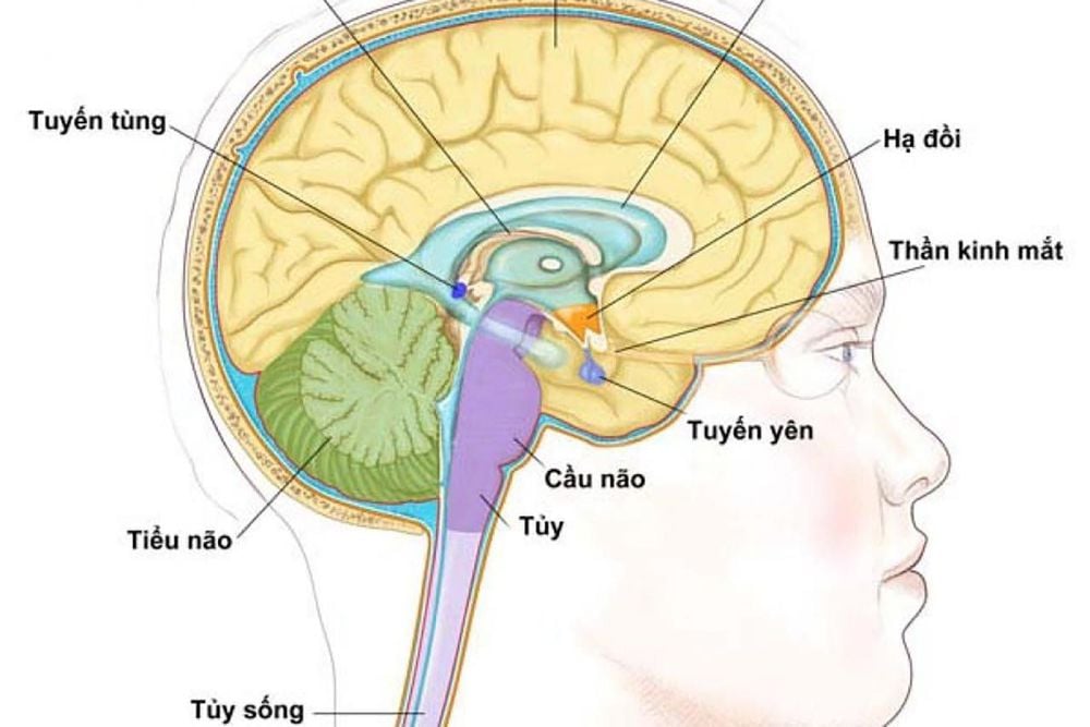
Tuyến yên là cơ quan đóng vai trò quan trọng không thể thay thế
2. Indications/contraindications for pituitary magnetic resonance imaging?
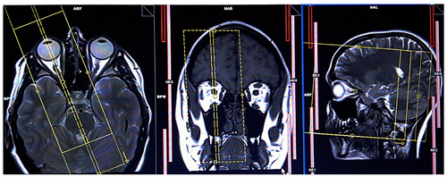
Pituitary magnetic resonance imaging is a safe and non-invasive imaging method. The purpose of pituitary magnetic resonance imaging is to detect local primary abnormalities or damage to adjacent areas affecting the pituitary gland. Indications for pituitary magnetic resonance imaging include:
Patients with suspected symptoms such as visual impairment, double vision, drooping eyelids, ocular paralysis, ... Patients with clinical manifestations by symptoms Symptoms suggest an increase or decrease in other hormones. The patient came to the clinic because of infertility, no abnormalities were detected in the genital organs. Presence of persistent headache, nonspecific Growth disturbance such as giant or dwarfism Pituitary tumor suspected. The patient has detected a tumor in the pituitary gland before, now it is necessary to monitor the development or evaluate the effect after treatment Menstrual disorders in female patients in the direction of amenorrhea, amenorrhea Encounter hypoglycemia Repeated repetition Changes in facial appearance and sensation Rapid weight gain, obesity Electrolyte disturbances Similar to other techniques, pituitary magnetic resonance imaging, although noninvasive and safe, cannot be used for all cases. The risks of magnetic resonance imaging are related to its magnetic field-dependent mechanism of action. Contraindications to pituitary magnetic resonance imaging include:
People with alternative devices or materials inside the body made from magnetic metal such as certain types of pacemakers, artificial heart valves, machines defibrillation, cochlear implantation, hemostasis clip, means of bone fusion,... People who are afraid of the dark or have claustrophobia. People carrying metal foreign bodies in the body such as shrapnel, especially in the two orbits and nearby areas. Young children with ADHD or those who are too young to lie still for long in one position. People who bring personal items into the shooting room such as jewelry, watches, bank cards, ...
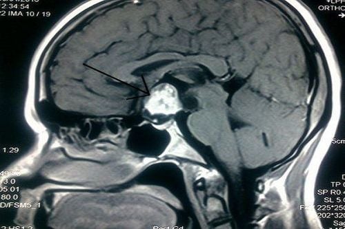
Bệnh nhân nghi ngờ u tuyến yên cần chụp MRI
3. Advantages of pituitary magnetic resonance imaging
Magnetic resonance imaging of the pituitary gland is increasingly being indicated in clinical situations. Compared with other imaging methods, pituitary magnetic resonance imaging has many outstanding advantages such as:
Patients are not exposed to radiation. The working mechanism of the magnetic resonance machine is based on the magnetic field. Therefore, pituitary magnetic resonance is considered quite safe, can be used for children and pregnant women. Contrast used in pituitary magnetic resonance imaging is derived from gadolinium, has a lower rate of allergic reactions or drug shock than iodinated contrast in computed tomography. Detecting small-sized lesions in the pituitary gland thanks to its ability to create detailed, high-contrast images. The image of the pituitary gland obtained by magnetic resonance imaging of the pituitary gland is many times clearer than that of cranial magnetic resonance imaging. In addition, the lesions in the brain parenchyma, if present, were also detected without being missed.
4. Notes before, during and after pituitary magnetic resonance imaging
4.1 Before shooting
Patients should keep the following notes in mind before taking a pituitary MR scan:
Cooperate when collecting information related to history to rule out contraindications to MRI. Do not bring personal items made from metal such as jewelry, watches, bank cards into the shooting room. Sign the commitment paper if you agree to perform the technique. Change clothes according to regulations, should wear earplugs or noise-reducing headphones.
4.2 While shooting
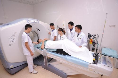
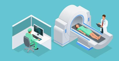
The patient needs to maintain a supine position on the imaging chair during the MRI scan. Keep the head position fixed in the magnetic field of the machine to avoid image noise. Don't worry or get nervous, try to stay calm and relaxed. Follow the instructions of the machine room technician. If there is an abnormality or discomfort during the scan, the patient should notify the medical staff.
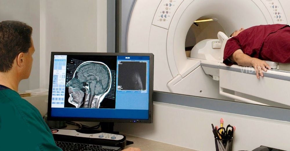
TRong quá trình chụp MRi nếu thấy biểu hiện bất thường cần báo cho nhân viên y tế
4.3 After shooting
If the pituitary magnetic resonance imaging procedure uses contrast, the patient will be monitored after the scan for at least 15 minutes. The patient then needs to rest and wait for about 30 minutes for the results to be interpreted. Vinmec International General Hospital with a system of modern facilities, medical equipment and a team of experts and doctors with many years of experience in neurological examination and treatment, patients can completely rest in peace. Center for examination and treatment at the Hospital.
Master, Doctor Trinh Van Dong has nearly 10 years of experience in the specialty of Diagnostic Imaging, especially has strengths in performing techniques: X-ray, ultrasound, computed tomography, magnetic resonance. Currently, Dr. Dong is working and working at the Department of Diagnostic Imaging, Vinmec Ha Long International General Hospital.
To register for examination and treatment at Vinmec International General Hospital, you can contact Vinmec Health System nationwide, or register online HERE.
MORE
Are pituitary tumors dangerous? Does magnetic resonance imaging (MRI) have any effect? Magnetic resonance imaging of the pituitary gland with injection of magnetic contrast (kinetic study) Dynamic


