
This is an automatically translated article.
The article is made by Master, Doctor Ton Nu Tra My - Department of Diagnostic Imaging - Vinmec Central Park International General Hospital.
Cranial magnetic resonance imaging with contrast injection is indicated in some special cases when it is not possible to evaluate the lesion or to suspect the lesion, which cannot be obtained by cranial magnetic resonance imaging without injection of contrast agent. detectable. This is a modern imaging technique, with high sensitivity and specificity, so it is very valuable in the diagnosis of neurological diseases.
1. Learn about the method of cranial magnetic resonance imaging with injection of magnetic contrast medicine
Contrast-injected cranial magnetic resonance (MRI) is a method of using contrast media to increase the contrast of anatomical structures that are normally difficult to see or distinguish. with surrounding structures, then using magnetic fields and radio waves to produce clear and detailed and clear cross-sectional images of the skull, especially brain tumors, thus helping to evaluate and detect lesions. injuries, abnormalities of the skull very early. Contrast drugs in diagnostic imaging help to accurately diagnose pathologies, with clarity and detail, especially tumors. In addition to the benefits that contrast agents bring, there can be complications related to contrast agents from mild to severe, the most severe is anaphylaxis, the patient can die quickly. However, this rate is very rare, it is necessary to equip an anti-shock medicine box at the imaging room. In which cases is cranial magnetic resonance imaging with injection of magnetic contrast agent indicated? Suspected brain tumor (including all brain tumors). Patients with persistent headaches that do not respond to analgesics, have signs of focal nerve paralysis, nausea, vomiting, loss of balance, ... can't find the cause of the headache, need to conduct resonance imaging. Cranium Suspected encephalitis, meningoencephalitis, brain abscess, brain fluke, .. the patient has symptoms of headache, fever, vomiting, clinical examination shows signs of stiff neck. Detection of cerebrovascular malformations. Epilepsy Cerebral stroke: Cerebral infarction, especially early cerebral infarction, cerebral bleeding in stages, arteriovenous infarction. In particular, it is possible to inject drugs in the case of using a perfusion pulse sequence
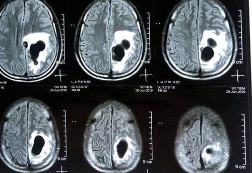
Hình ảnh người bệnh mắc sán não được phát hiện qua MRI
2. Cranial magnetic resonance imaging procedure with magnetic contrast injection
Prepare:
Medicine:
Sedative for people who cannot lie still like children, stimulated patients Contrast drug from Antiseptic solution, cotton Anti-shock medicine box and first aid equipment for drug accident Other supplies: needle, syringe, gloves Patient:
The patient is fully explained about the technique, purpose, and complications caused by contrast, agrees to sign the commitment to take MRI brain with contrast injection The patient is instructed to change clothes, remove jewelry, bronze, metal on the body The patient does not need to fast, instruct the patient to lie still. suspect

Người bệnh được giải thích đầy đủ về kỹ thuật chụp cộng hưởng từ sọ não có tiêm thuốc đối quang từ trước khi thực hiện
Steps to perform
Step 1: Move the patient into the machine compartment. The patient is gently moved onto the imaging table, the patient lies in the supine position, then moves the table into the machine compartment
Step 2: Control the scanner from the control room
Capture position Select diagnostic pulse sequences appropriate for the purpose of the examination. Perform normal pulse sequences: T1, T2, Flair for all subjects in various cutting directions including horizontal (horizontal), horizontal (horizontal) and vertical (vertical). Select special pulse sequences for the pathology to look for such as diffusion pulse series for brain injury related lesions, brain tumor, brain abscess, T2* pulse sequence to find bleeding brain lesions,. .. Step 3: Process the image. The technician runs each pulse and processes the images obtained on the workstation screen, selecting the clearest images needed to reveal the pathology for film printing.
Step 4: Inject the contrast agent.
Conduct pulse localization for drug injection (possibly T1 pulse cuts in 3 directions), then inject contrast agent intravenously and run selected pulses. Step 5 . The image is processed, then the doctor reads the lesion, describes it on the computer, concludes and prints the results. The displayed images must ensure to clearly show the anatomical structures in the examination area, detect lesions, evaluate the nature of enhancement.

Hệ thống máy chụp cộng hưởng từ tại Bệnh viện Đa khoa Quốc tế Vinmec
Vinmec International General Hospital is one of the hospitals that not only ensures professional quality with a team of leading medical doctors, modern equipment and technology, but also stands out for its examination and consultation services. comprehensive and professional medical consultation and treatment; civilized, polite, safe and sterile medical examination and treatment space. Customers when choosing to perform tests here can be completely assured of the accuracy of test results.
Master. Dr. Ton Nu Tra My is currently a Doctor of Radiology, Vinmec Central Park International General Hospital. Dr. Tra My used to be a lecturer in the Department of Diagnostic Imaging, Hue University of Medicine and Pharmacy and had a long time working at the Department of Diagnostic Imaging, Hue University of Medicine and Pharmacy Hospital.
To register for examination and treatment at Vinmec International General Hospital, customers can call Hotlines of hospitals or register online HERE.