This is an automatically translated article.
This article is professionally consulted by Master, Resident Doctor Tran Duc Tuan - Department of Diagnostic Imaging - Vinmec Central Park International General Hospital.Magnetic resonance imaging (MRI) of the thoracic spine is an imaging procedure that examines the entire complex structure of the thoracic spine. Unlike X-rays or computed tomography (CT scans), MRI does not use ionizing radiation, reducing the risk of radiation exposure for the patient, but still maintaining image quality and supporting the procedure. diagnosis of diseases in this area.
1. Anatomy of the thoracic spine
The spine is made up of 33 vertebrae that are separated by intervertebral discs and divided into distinct areas.Neck segment: consists of 7 vertebrae in the neck. Thoracic segment: includes 12 thoracic vertebrae. Lumbar segment: consists of 5 vertebrae in the lower back. Sacral segment: 5 small vertebrae fuse together. Trunk: 4 remaining vertebrae, fused to form 1 coccyx The spinal cord, a major part of the central nervous system, is located in the vertebral canal. The function of the spinal cord is to carry sensory and motor signals from the brain and control many reflexes.
The thoracic vertebrae and related components, the spinal cord, and the nerve roots that emerge from this segment are the target of MR imaging of the thoracic spine.
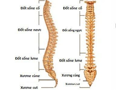
Cột sống được tạo thành từ 33 đốt sống
2. What are the reasons to have an MRI of the thoracic spine?
Magnetic resonance imaging of the thoracic spine can be used to examine the bone or the inner spinal cord, for damage or the presence of structural abnormalities, or for certain other conditions, such as:
Tumor Abscess Congenital abnormalities Aneurysms Venous malformations Hemorrhage, or bleeding into the spinal cord Subdural hematoma around the spinal cord Degenerative spondylosis, multiple sclerosis, spinal cord disease Herniated disc or degenerative disc disease of the spinal cord Help plan surgery on the spine, such as pinched nerve decompression or spinal fusion.

Chụp cộng hưởng từ cột sống ngực có thể được sử dụng để kiểm tra phần xương
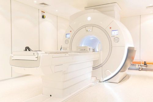
3. Things to know about the thoracic spine magnetic resonance imaging procedure without injecting magnetic contrast
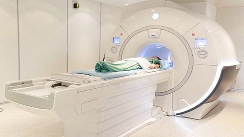
Eat / Drink : You can eat, drink and take your medicine as usual. Clothing: You must completely change into a specialized gown and remove all personal belongings, especially metal objects. Posture: You are placed on the slider into the large magnet cage and need to lie completely still for a sharp, quality image. Due to the loud noise of the MRI machine during operation, you may be given earplugs to avoid noise. Avoid Anxiety: If you need anti-anxiety medication because of your fear of being surrounded, talk to your doctor directly. At the same time, after the MRI scan is over, you can't go home yet, but you need to stay in the hospital for monitoring until the drug's effect expires and you will also need your loved one to take you home. Contraindications: In general, there is no problem with MRI except in cases where there are permanent metal objects and machines on the body, eg pacemakers, pacemakers. If you are pregnant or think you may be pregnant, discuss this with the doctor who ordered the MRI. Scanning process: The time to perform magnetic resonance imaging of the thoracic spine without injection of contrast agent usually lasts for 30 minutes. The technician will be in another room where the scanner controller is located. However, you are always under constant surveillance. Speakers inside the scanner will allow the technician to communicate and listen to you. You'll have a call button so you can let the technician know if you have any issues along the way. A technician will monitor you and will be in regular contact if there are any issues to ask. It is important to remember that you must remain in the position throughout the exercise. Any movement can cause distortion and affect the quality of the scanned image. After the scan: Once the scan is complete, the table will slide out of the scanner and you will be assisted out of the table. Although the MRI procedure is painless, having to lie still for the duration of the procedure can cause certain discomfort, especially in the case of recent trauma or invasive procedures such as surgery. The technician will use all measures to make you as comfortable as possible and complete the procedure as quickly as possible to minimize any discomfort or pain. At the end of the scan: You should move slowly when getting up from the scanner table to avoid any feeling of dizziness from having to lie flat for a long time of the procedure. If any sedation has been taken, you will be asked to rest until the sedation wears off and you will also need to avoid driving or operating machinery for at least a day afterwards. When you get home, you can completely return to your normal activities. The results of thoracic spine magnetic resonance imaging without contrast injection will be verified by an imaging specialist before being returned to the appointing doctor. By incorporating additional clinical findings as well as other diagnostic means, thoracic spine magnetic resonance imaging will enable diagnostic support as well as subsequent treatment modalities.
In summary, magnetic resonance imaging without injection is a useful diagnostic imaging tool to investigate problems in the thoracic spine in particular and problems related to bones, joints and nerves in general. However, for the best quality of collection, patients need to know and strictly follow the regulations in the magnetic resonance imaging procedure as above.
Vinmec International General Hospital is one of the hospitals that not only ensures professional quality with a team of leading doctors, modern equipment and technology, but also stands out for its examination and consulting services. and comprehensive, professional medical treatment; civilized, polite, safe and sterile medical examination and treatment space. Customers when choosing to perform tests here can be completely assured of the accuracy of test results.
Master, Doctor Tran Duc Tuan is well-trained in the country and many centers have prestigious medical background in the world such as: Australia, Singapore, Thailand... The doctor has many years of experience in the field of medicine. imaging and endovascular and extravascular interventions. Currently, the doctor is working at the Department of Diagnostic Imaging and Interventional Vascular Unit - Vinmec Central Park International General Hospital.
Please dial HOTLINE for more information or register for an appointment HERE. Download MyVinmec app to make appointments faster and to manage your bookings easily.