This is an automatically translated article.
The article is professionally consulted by MSc, BS. Dang Manh Cuong - Radiologist - Radiology Department - Vinmec Central Park International General Hospital. The doctor has over 18 years of experience in the field of ultrasound - diagnostic imaging.Magnetic resonance imaging of the thoracic spine with magnetic contrast injection is a modern and useful diagnostic imaging tool, providing clear images of anatomical structures in the thoracic spine in particular and the thoracic region in particular. shared. This is the basis for supporting diagnosis for diseases in the thoracic spine as well as surgical intervention if indicated.
1. What is thoracic spine magnetic resonance imaging with injection of magnetic contrast?
Magnetic Contrast Magnetic Resonance Imaging (MRI) of the thoracic spine uses strong magnets, radio waves, and a computer to create very clear and detailed images of your spine.Anatomical components in this area include the spine, discs, ligaments, spinal cord and nerve roots and surrounding soft tissue. These organs are often difficult to examine with universal imaging tools such as X-ray or computed tomography, because of the presence of both hard and soft tissue. Therefore, with magnetic resonance imaging, the thoracic spine area will clearly show every detail, helping to define the disease more accurately.
Besides, with the participation of the contrast agent, the perfused soft tissues will appear better. The injection periods will reflect the local pathophysiology, especially useful in inflammatory diseases or neoplasms.

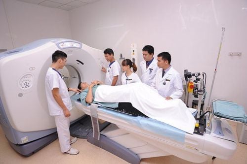
Chụp cộng hưởng từ, vùng cột sống ngực sẽ hiện lên rõ ràng từng chi tiết, giúp việc phân định bệnh được chính xác hơn
2. What are the indications for magnetic resonance imaging of the thoracic spine with magnetic contrast injection?
Your doctor will need to take a thoracic spine with contrast injection since you have health problems in the spine such as back pain, neck pain that radiates down the back, numbness, tingling in the arms or weakness in the arms. hand.Now, an MRI allows your doctor to examine the small bones, called vertebrae, that make up your spine, as well as the spinal discs, spinal canal, and spinal cord. Through it, the doctor can diagnose diseases in the spine such as:
Deformities or abnormal curvature in the spine Vertebral fractures Spinal trauma Spinal infections Spondylitis inflammation degenerative disc herniation spinal cushion Compression tumor Although an MRI can scan your entire spine, with the conditions listed above, a thoracic spine MRI may be sufficient for diagnosis. However, unlike the X-ray scans used in X-rays and computed tomography, magnetic resonance imaging does not use radiation, so it does not cause any harm.
In addition, your doctor may also order MRI of the spine to help plan surgeries on the spine, such as decompressing a pinched nerve or needed for procedures such as an epidural or steroid injection.
3. Magnetic resonance imaging procedure of thoracic spine with injection of magnetic contrast agent
Because MR imaging of the thoracic spine involves an injection of magnetic contrast, you will be asked to fast for at least 6 hours prior to the scan. However, taking medicine to treat chronic diseases still needs to be done regularly every day, even in the morning before going for magnetic resonance imaging.When you come to the imaging room, you need to change into a specialized gown that is to remove metal objects on the body, including eyeglasses, hearing aids, jewelry, watches and other items. In addition, tell your doctor if you have any of the following:
Have serious medical conditions, such as kidney or liver failure Have had recent surgery Have any allergies or asthma Are pregnant or think you may be pregnant Have metal objects or electronic devices that can affect the MRI images, including: Prosthetic heart valves, prostheses, or artificial joint replacements, metal beads for adjustment treatment for brain aneurysms, cochlear implants, implantable pumps for continuous injection of drugs such as insulin, metal fragments, such as bullets or shrapnel, batteries, screws, steel plates, stents or surgical steel threads surgery, pacemaker or defibrillator. If such items are not removable, the patient will be considered contraindicated and cannot undergo magnetic resonance imaging and need to be replaced with other imaging facilities.
Furthermore, if you have a tattoo or permanent makeup, some tattoo inks contain iron or metals in general, which may heat up during magnetic resonance imaging.
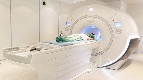
When everything is ready, you will be arranged to lie on a table that slides into the body, which is essentially a giant magnet cage. If you don't like being confined in a small space or you are too nervous or stressed, let your doctor know and a sedative to help you relax will be prescribed.
The technician will determine the area to be scanned is the thoracic spine. The first scan is called magnetic resonance imaging of the thoracic spine without contrast injection.
Then a certain amount of contrast agent is injected into a vein in the arm or on the back of the hand. This medicine will help your doctor see more clearly any infection, tumor, or disc damage in your spine. The dye commonly used in MRI is called gadolinium. When injected into the body, a small number of people may feel flushed or cold for a few minutes afterward, but only briefly.
During the entire time you lie on the table that slides into the MRI machine, you will need a strap to help you stay in place during the examination. Also, there is always a radiologist and technician outside the room, but they can still see, hear and talk to you at any time. Sometimes there may even be a relative who can be in the room with you, helping to relieve your stress.
When working, the MRI machine will create a strong magnetic energy around you. The computer then takes the signal from the MRI and uses it to create a series of images, each showing a thin slice of your body. Furthermore, magnetic resonance imaging can create images in three-dimensional space, making it easier to locate lesions.
Usually, each image slice is a single scan of the body by the MRI scanner probe. Each run of such a machine can last from a few seconds to several minutes. However, you should always keep your whole body still during the scan, which can take anywhere from 30 minutes to an hour or more, depending on the extent of the slice of your spine.
You will feel absolutely no pain during the MRI, but you may feel warmth in the area of your spine during the scan. In addition, you will also hear very noisy operation when images are recorded. At this point, earplugs or headphones can help block out the noise if it's uncomfortable, or you can even listen to music instead.
4. How to monitor after magnetic resonance imaging of the thoracic spine with magnetic contrast injection
Once the MRI of the spine is finished, you can return to your normal activities immediately. However, if you need a sedative to relax you before the scan, you'll need to stay in the hospital for observation until the drug's effects are over. After that, you also need to have a loved one to take you home and absolutely not use any vehicle or machine for at least 24 hours afterwards.Sometimes contrast injection can cause side effects, although the incidence is very low compared to contrast in computed tomography. At this point, you may feel nauseous, have a headache, or you may have pain at the site where the dye was injected. However, an allergic reaction to the dye is very rare, but if you experience hives, itchy skin, itchy eyes or any other symptoms that suggest an allergy, tell your doctor right away. to be dealt with in a timely manner.
The time required to analyze MRI images of the thoracic spine with contrast injection is usually very long and you do not need to wait for the results.
A radiologist will read the results and send them back directly to the referring doctor. Combined with the clinical symptoms and other tests, you will be scheduled to respond with the results of the X-ray and follow-up treatment within a few days.
In case you need surgical intervention, the results of this magnetic resonance imaging should be stored carefully because it will be the basis for comparing and contrasting the effectiveness of surgery later.
In conclusion, contrast-enhanced thoracic spine magnetic resonance imaging today seems to have replaced most other imaging modalities when investigating in this area. With clear, sharp images, clearly distinguishing structures, patients can find diseases quickly and accurately and intervene more effectively, improving quality of life.

Tiêm thuốc đối quang từ có thể gây ra tác dụng phụ, mặc dù tỷ lệ xảy ra rất thấp so với thuốc cản quang trong chụp cắt lớp vi tính
Please dial HOTLINE for more information or register for an appointment HERE. Download MyVinmec app to make appointments faster and to manage your bookings easily.