This is an automatically translated article.
Long QT syndrome is a disorder of the heart's electrical system. Torsade de pointes (TdP), as indicated on the electrocardiogram (ECG), is a feature of ventricular hyperrhythmias that present complex torsades de pointes. The QT interval is the upper part of the electrocardiogram, representing the time it takes for the electrical system to fire impulses through the ventricles and then recharge.
1. What is long QT syndrome and torsades de pointes?
Long QT syndrome occurs due to defects in ion channels, causing a delay in the time the electrical system recharges with each heartbeat. When the QT interval is longer than normal, it increases the risk of torsades de pointes, a life-threatening form of ventricular tachycardia. The electrical activity of the heart is generated by the flow of ions (charged particles of sodium, calcium, potassium, and chloride) into and out of the heart's cells. Microscopic ion channels control this flow. This condition may not seem worrisome at first, but in the long run it can lead to serious health problems. Drugs that prolong the QT interval are one of the causes of long QT syndrome. However, this does not happen often.
Torsade de pointes is an uncommon and distinctive form of polymorphic ventricular tachycardia (VT), characterized by a gradual change in amplitude and twisting of the QRS complex around the isoelectric line (see image below) . Torsade de pointes is associated with a prolonged QT interval, which may be congenital or acquired. Torsion usually ceases spontaneously but frequently recurs and can degenerate into ventricular fibrillation.
In torsades de pointes, the morphology of the QRS complex changes with each beat. The ventricular rate can range from 150 beats per minute (bpm) to 250 bpm.
The initial report describes the frequent change in the morphology of the QRS vector from positive to negative and back again. Most cases exhibit polymorphism, but the axial shift may not have regularity.
The definition also requires a marked increase in the QT interval (usually ≥600 msec). Cases of polymorphic ventricular tachycardia in which the QT interval is not prolonged are treated as generalized ventricular tachycardia. The torsion usually occurs in waves that do not last long. Thus, the rhythm band usually shows the patient's baseline QT prolongation.
The cause and management of torsion is generally quite different from that of garden VT. In particular, the use of class IA antiarrhythmic drugs, which tend to prolong the QT interval, has the deleterious consequence of torsion. Therefore, it is extremely important to distinguish between these entities

Hình ảnh so sánh hội chứng QT dài và điện tim bình thường
2. Pathophysiology of congenital and long QT syndrome
The link between torsion and QT prolongation has been known for a long time, but the mechanisms involved at the cellular and ionic levels have been elucidated over the past decade or so. The underlying abnormality of both acquired and congenital long QT syndrome is in the ion flux during repolarization, which affects the QT interval.
A variety of changes in ion current can cause the general effect of decreased repolarizing current, reflected in a long QT. These changes can lead to secondarily the following depolarization currents and sometimes action potentials, called afterdepolarizations. This leads to a further delay in repolarization and induces early post-depolarization (EAD), the event that causes torsion. Repolarization has 3 stages.
Stage 1: During the initial activation of the action potential in normal cardiac cells, a net rapid influx of positive ions (Na + and Ca ++ ) occurs, leading to depolarization of the heart cell. membrane. This is followed by an extremely fast, transient potassium current (Ito), while the flow rate of positive ions (Na + , Ca ++ ) decreases. This represents the initial part of the repolarization process. Stage 2 is characterized by plateaus. The positive currents flowing in and out become almost equal during this period. Stage 3 of repolarization is accomplished by triggering a delayed rectifier potassium (IK) current that moves outward while the positive current is directed inward to decay. If slow inactivation of the Ca++ and Na+ currents occurs, the inward current can cause single or repeated depolarization in stages 2 and 3 (i.e. EAD). These EADs appear as pathological U waves on the surface electrocardiogram, and when thresholded, they can cause ventricular tachyarrhythmias. These repolarization changes do not occur in all cardiomyocytes. The deep endocardium and middle myocardium (including M cells) of the ventricles are more susceptible to repolarization and prolonged EAD because they have a less rapid rectified potassium current (IKr). While other regions may have short or normal cycles. Heterogeneity of repolarization in cardiomyocytes promotes the propagation of activated activity, which is initiated by EADs by a recirculation mechanism and is now thought to be responsible for the maintenance of repolarization. twisted.
Six basic genetic variants are currently recognized. Genotypes LQT1 and LQT2 have slow potassium channels, while LQT3 shows defects in sodium channels. The modality of early treatment may be based on the individual's genotype.
3. Causes of congenital and long QT syndrome
QT interval prolongation can be congenital, as seen in Jervell and Lange-Nielsen syndrome (i.e. congenital long QT associated with congenital deafness) and Romano Ward syndrome (i.e. separate prolongation of the QT interval). ). Both of these syndromes are associated with sudden death from primary ventricular fibrillation or from the degeneration of torsades de pointes into ventricular fibrillation.
Electrolyte disturbances that have been reported to cause torsion include hypokalemia. These disorders cause a delay in stage III (ie, prolongation) and form the basis for the occurrence of arrhythmias. Close observation is required in predisposed patients, such as those with cirrhosis or hypothyroidism.
Risk factors
Risk factors for torsades de pointes include:
Congenital long QT syndrome Female gender Acquired long QT syndrome (causes include medications and electrolyte disturbances such as hypokalemia and hypokalemia) Bradycardia Basic ECG abnormalities Renal or hepatic failure Congenital (adrenergic dependent) long QT syndrome
The following congenital syndromes are associated with torsion:
Jervell's syndrome and Lange-Nielsen Romano-Ward syndrome Acquired long QT syndrome
Drugs in several drug classes have been associated with torsion.
Antiarrhythmics associated with torsion include:
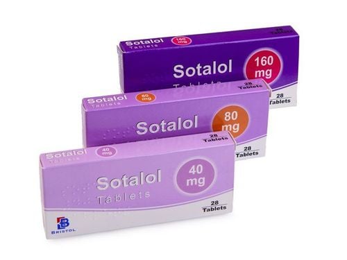
Class IA - Quinidine, disopyramide, procainamide Class III - Sotalol, amiodarone (rare), ibutilide, dofetilide, almokalant Other classes of drugs associated with torsades de pointes include :
Antibiotics - Erythromycin, clarithromycin, azithromycin, levofloxacin, moxifloxacin, gatifloxacin, trimethoprim-sulfamethoxazole, clindamycin, pentamidine, chloroquine Antifungals - Ketoconazole , itraconazole Antiviral drugs - Amantadine Antipsychotics - Haloperiazines, phenothiazines thioridazine, trifluoperazine, sertindole, zimeldine, ziprasidone Tricyclic and quadricyclic antidepressants Antihistamines (histamine receptor antagonists 1) - Terfenadine, astemizole, diphenhydramine, hydroxyzine Cholinergic antagonists - Cisaprid, organophosphates (insecticides) ) Diuretics - Indapamide, hydrochlorothiazide, furosemide Antihypertensives - Bepridil, lidoflazine, prenylamine, ketanserin Lithium Anticonvulsants - phenytoin, carbamazepine (possibly) Oral hypoglycemic Citrate (massive blood transfusion) Cocaine Vasopressin (possibly) Fluoxetine (possibly) Anesthesia - dexmedetomidine, propofol Loperamide - This antidiarrheal has been linked to torsion, even in patients with no genetic or cardiac defects, often due to abuse or misuse.

Thuốc kháng vi-rút – Amantadine là một trong các nguyên nhân hội chứng QT dài
Some drugs (eg, amiodarone) commonly prolong QT but have little relevance to the clinical consequences of long QT.
Conditions associated with torsion include the following:
Electrolyte abnormalities - Hypokalemia, hypokalemia, hypocalcemia Endocrine disorders - Hypothyroidism, hyperparathyroidism, pheochromocytoma, hyperaldosteronism, hypoglycemia Cardiac condition - Myocardial ischemia, myocardial infarction, myocarditis, arrhythmia, complete atrioventricular (AV) block, takotsubo cardiomyopathy Intracranial disorders - Subarachnoid hemorrhage, thalamic hematoma, cerebrovascular accident, encephalitis, head trauma Nutritional disorders - Anorexia nervosa, hunger, liquid protein diet, gastric dilatation.
4. Differential diagnosis of torsades de pointes
The differential diagnosis of torsades de pointes includes the following:
Pediatric ventricular tachycardia, Tachycardia Syncope Renal failure, chronic and dialysis complications Toxicity, antiarrhythmic Toxicity, antihistamine Vibration Sudden cardiac death Another consideration is to distinguish acquired long QT syndrome from congenital long QT syndrome. In addition, torsion should be differentiated from polymorphic ventricular tachycardia or, rarely, monomorphic ventricular tachycardia.
Supraventricular tachycardia with abnormal conduction can be confused with torsion, especially when the degree of deviation varies. A previous clue was that atrial fibrillation could be mixed with narrower and typical QRS complexes.
Regular ECG monitoring is indicated for patients at risk due to chronic conditions or drug therapy. When the patient is in sinus rhythm, check the QT interval. Typically, the QT interval is prolonged and a pathological U wave is present, reflecting abnormal ventricular repolarization. The most consistent indicator of QT prolongation is a QT of 0.60 s or more or a QTc (corrected for heart rate) of 0.45 s or more.
Other ECG features useful in diagnosing torsion include the typical mode of onset and its morphology, as follows:
Patients have exacerbations of 5-20 beats at a faster rate 200 bpm; Occasional sustained episodes can be seen Gradual change in polarity of QRS to isoelectric occurs Complete 180° torsion of QRS complex in 10-12 beats is present A short-long sequence- The short interval between RR intervals occurs before the activation response. Patients may recover spontaneously or switch to non-polymorphic ventricular tachycardia or ventricular fibrillation. Alternate T waves can sometimes be seen before torsion. must be preceded by a pause in most cases. In congenital long QT syndrome (adrenergic dependence), transient dependence is found in most cases of adults, whereas torsades de pointes are not transiently dependent in children.
This rhythm failure can happen for a variety of reasons. During very short torsion runs, the typical twisting of the QRS complex around the isoelectric line may not be obvious. Initial events are often short-lived. In the case of single-lead recording, the typical morphology of the torsion may not be obvious.
The diagnosis of torsion should be considered in any patient with pause-dependent ventricular tachycardia, and ventricular size in patients with a long QT interval may be indicative of impending torsades de pointes.

Ngất là một trong các yếu tố chẩn đoán phân biệt của xoắn đỉnh
5. Torsion treatment
5.1. Acute management Treatment can be divided into short-term and long-term management.
5.1.1. Short-term management of torsades de pointes. In case of prolonged torsades de pointes and does not go away on its own after 5 seconds, the following treatment methods will be applied:
Punch in front of the heart, press the heart outside the chest. Intravenous infusion of 1.5 - 3 grams of magnesium sulfate Discontinue current antiarrhythmic drugs (class Ia, Ic, III antiarrhythmic drugs, tricyclic antidepressants, some H1 antagonists, erythromycin... ). Do not give electric shock. If the torsades de pointes are prolonged, the risk of onset of ventricular fibrillation will be applied with the following treatments:
Punch to the pre-cardiac region 200 J extrathoracic shock (avoid repeated shocks that can cause potassium loss) Adjust blood potassium: maintain blood potassium 4-5mmol/l by infusion of potassium chloride 0.5-1g/1h (electrical pump) for 3 hours and then IV infusion of 6g/24 hours Combine IV infusion 1 -3 grams of magnesium sulphate for 2 hours and then continue IV infusion 6-12g/24 hours Place a pacemaker with electrodes in the heart chamber, set the frequency to 100-120 if torsades de pointes recur, potassium disturbances have not been corrected If due to atrioventricular block, IV isoprenaline 1-2 mg or 0.5 mg IV atropine while waiting for pacemaker insertion. In another stable patient, direct current (DC) reduction in heart rate was maintained as a measure of last resort, because torsion is paroxysmal and is characterized by frequent recurrences after attenuation. heartbeat. Although torsion frequently ends on its own, it can degenerate into ventricular fibrillation, requiring DC defibrillation.
Any offending agent must be withdrawn. Predisposing conditions such as hypokalemia, hypokalemia, and bradycardia should be identified and corrected.
5.2. Pharmacological therapy 5.2.1. Magnesium Magnesium is the drug of choice to prevent premature arrhythmias (EADs) and terminate arrhythmias. Magnesium achieves this by reducing calcium intake, thereby reducing the amplitude of the EAD.
Magnesium can be injected initially at a dose of 1-2 g IV over 30-60 seconds, then can be repeated after 5-15 minutes. Alternatively, a continuous infusion can be initiated at a rate of 3-10 mg/min. Magnesium is effective even in patients with normal magnesium levels. Because of the danger of hypermagnesaemia (impaired neuromuscular function), the patient should be closely monitored.
Some authorities recommend potassium supplements to raise potassium levels to high-normal levels, which increases the outflow of potassium from cardiomyocytes, thereby causing rapid repolarization.
5.2.2. Lidocaine Lidocaine is usually ineffective during torsion. Sometimes it may have an initial beneficial effect, but the torsion repeats in all cases.
Mexiletine may also be helpful in preventing torsion. In one study, it was used in HIV-infected patients with long QT interval and torsion. It effectively prevents torsion on a long term basis.
Patients with congenital long QT syndrome are thought to have balance or sympathetic tone abnormalities and are treated with beta-blockers. If the patient has a breakthrough torsion, a short-acting beta-blocker, such as esmolol, can be tried.

Lidocain là một loại thuốc thường không có tác dụng với điều trị xoắn đỉnh
5.2.3.Isoproterenol Isoproterenol can be used in cases of bradycardia-dependent torsion commonly associated with acquired long QT syndrome (pause-dependent). It should be given as a continuous intravenous infusion to keep the heart rate above 90 bpm.
Isoproterenol increases AV conduction rate and decreases QT interval by increasing heart rate and decreasing temporal dispersion of repolarization. Beta-adrenergic agonists such as isoproterenol are contraindicated in the congenital form of long QT syndrome (adrenergic dependent). Due to the precautions, contraindications, and side effects associated with its use, this drug is used as a temporary agent until excessive acceleration may begin.
5.3. Temporary Intravenous Infusion Rate Based on the fact that the QT interval shortens with a faster heart rate, bradycardia can be effective in terminating torsion. It is effective in both forms of long QT syndrome because it facilitates repolarization of potassium currents and prevents long pauses, inhibits EAD, and reduces the QT interval.
Atrial pacing is the preferred modality as it preserves the atrial contribution to ventricular filling and also results in a narrower QRS complex and thus a shorter QT. In patients with AV block, ventricular pacing can be used to inhibit torsion. This depends on intact atrioventricular conduction at the required pacing rate.
The pacing should be set at 90-110 bpm until the QT interval is normalized. Overruns may be necessary with speeds up to 140 bpm for rhythm control.
Patients with extreme stage torsion should be treated with defibrillation or electrical defibrillation. Anecdotal reports cite successful conversion to phenytoin (Dilantin) and lidocaine. Several cases of successful conversion using phenytoin and speed-up have been reported.
If the patient does not respond to conversion to phenytoin and pacing is too fast, try electrical cardioversion.
5.4. Long-term treatment Beta-adrenergic antagonists at maximally tolerated doses are used as first-line long-term therapy in congenital long QT syndrome.
Propranolol is the most widely used, but other agents such as esmolol or nadolol may also be used. Beta-blockers should be avoided in congenital cases where bradycardia is a prominent feature. Beta-blockers are contraindicated in acquired long QT syndrome because the bradycardia produced by these agents can cause torsion. One approach to assess the adequacy of beta blockade is by exercise testing. One investigator recommends aiming to reduce the maximum heart rate by at least 20% from baseline (beta-blocker pretreatment). Another approach is to check blood levels of beta-blockers (eg, propranolol) when possible. Patients with no syncope, ventricular tachycardia, or family history of sudden cardiac death may be observed without initiating treatment.
Permanent pacing is beneficial for patients who remain symptomatic despite receiving maximally tolerated beta-blocker doses and can be used in addition to beta-blockers. It reduces the QT interval by enhancing the polarizing potassium current and blocking EAD.
High left thoracic sympathectomy, another antimycotic therapy, is effective in patients unable to take beta-blockers and pacing. Accidental severing of the sympathetic nerve in the eye can lead to Horner's syndrome.
Implantable defibrillators (ICDs) are useful in cases of recurrent torsion despite treatment with beta-blockers, pacing, and possibly left thoracic sympathectomy. Beta-blockers should be used in conjunction with an ICD because shock may further cause torsion due to adrenergic stimulation. In the United States, an ICD for refractory cases is often possible before sympathectomy.
Usually no long-term treatment is needed in acquired long QT syndrome because the QT interval returns to normal after the predisposing factor or predisposition has been corrected. Pacemaker implantation is effective in cases associated with heart block or bradycardia. ICDs are indicated in cases that cannot be managed by avoiding the offending agent.
The line between acquired and congenital may not always be clear. Secondary factors are often present, and individuals may show increased sensitivity to the effects of QT.
Complications Complications may include the following:
Monomorphic ventricular tachycardia Ventricular fibrillation Sudden cardiac death Prognosis In congenital long QT syndrome, mortality for untreated patients is 50% in 10 years, can be reduced to 3-4% with treatment intervention.
In acquired long QT syndrome, the prognosis is good when the provoking factor has been reliably identified and eliminated.
Note Use the drug only with the consent of the doctor. Instruct patients to avoid competitive sports (in the case of congenital long QT syndrome).
Close monitoring is required because of the risk of sudden cardiac death. Provide emotional support; suggest joining a cardiac support group.
Patients should be taught how to monitor their pulse and recognize drug side effects. Families should undergo counseling for basic life support.
Please dial HOTLINE for more information or register for an appointment HERE. Download MyVinmec app to make appointments faster and to manage your bookings easily.
References: msdmanuals, emedicine.medscape, ecgwaves


