This is an automatically translated article.
The article was written by Dr. Trinh Van Dong, Doctor of Radiology - Department of Diagnostic Imaging - Vinmec Ha Long International General HospitalDiagnosis of lower extremity artery disease can be based on 2D ultrasound and color doppler ultrasound to determine the location and morphology of the lesion and evaluate the hemodynamics of the flow at and after the lesion site.
1. What is lower extremity artery disease?
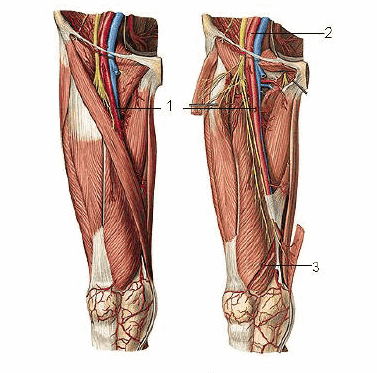
Lower extremity artery disease is a pathological condition of the iliac and lower extremity arteries in which the lumen of the artery is narrowed or blocked, resulting in reduced perfusion of the muscles and related downstream organs such as the skin and nerves. . The risk factors of lower extremity artery disease include smoking, diabetes, high blood pressure, high cholesterol... Nowadays, lower extremity artery disease tends to increase rapidly in number which can be due to disease Patients with metabolic diseases increase as well as the patient's nutritional regimen changes. Patients may present with symptoms such as pain, loss of pulse, pallor, paresthesia, loss of movement, coldness of the affected limb or no symptoms, however, in addition to the diagnosis based on clinical symptoms, it is also based on clinical symptoms. In the Ankle Brachial Index (ABI) at rest, if the ABI index is ≤ 0.9, it is diagnostic. Diagnosis of lower extremity artery disease can be based on 2D and color Doppler ultrasound to determine the location and morphology of the lesion and to evaluate the hemodynamics of the flow at and after the lesion site. However, to accurately assess the lesion, it is necessary to perform computed tomography of the lower extremity arteries with intravenous contrast injection.

Tiêm thuốc cản quang vào tĩnh mạch trước khi chụp
2. Why do computerized tomography arteries of the lower extremities?
Computerized tomography, also known as CT-Scan (Computer Tomography Scan), is a method of using X-rays. In addition to diagnosing lower extremity artery disease by clinical symptoms, 2D ultrasound and color doppler ultrasound, Diagnosis by multi-segment computed tomography with contrast injection of lower extremity arterial system and vascular reconstruction is extremely important and helps clinicians in making treatment decisions. for patients is medical treatment, surgery (endovascular removal, bypass surgery, stenting, balloon dilation or amputation). Contrast-enhanced lower extremity angiography allows assessment of the location, growth, localized or diffuse spread of the lesion, the development of collateral circulation, and the characteristics of the arteries. small circuit downstream. Contrast-enhanced lower extremity computed tomography angiography allows to avoid unnecessary complications associated with invasive angiography. Therefore, at present, multi-slice computed tomography of the lower extremities with contrast injection is one of the first priority techniques to diagnose and evaluate lower extremity artery disease.

Chụp cắt lớp vi tính là phương pháp chẩn đoán hình ảnh không xâm lấn
3. What is the procedure for computed tomography of the lower extremity arteries?
Computed tomography is a technique used in many large hospitals, especially at Vinmec General Hospital. It will be performed by technicians with many years of experience, must follow a strict process. and read the results by doctors who are leading experts in the field of diagnostic imaging. Below are the steps to perform a CT scan of the lower extremities: Examination of the patient: Before performing the imaging process, the patient will be clinically examined by the doctor, carefully asked about the medical history first. Thereafter, a history of allergies and mandatory tests such as liver function and kidney function should be performed. Prepare the patient and equipment: The patient will be clearly explained about the imaging procedure, about the patient's position. If the patient has a history of allergies or asthma, depending on the specific case, the patient will be treated for allergy prevention according to the specific procedure of Vinmec International General Hospital. Computerized tomography of the lower extremities requires contrast injection, so the technician must be prepared to place the intravenous needle, prepare the contrast agent, and the shock kit.

Bệnh nhân thực hiện chụp CT -Scan theo hướng dẫn của kỹ thuật viên
Perform CT-Scan: The patient lies on the table, moves to the center of the machine. An X-ray generator will pass through the part to be scanned and to the receiver on the opposite side. The patient will have thin cross-sections from the iliac aortic confluence to the sole of the foot two times before and after contrast injection. The data obtained from the receiving unit will be transmitted to the computer system that processes the image and produces the final result. Read and return results: After taking the picture, the radiologists will use image processing software, angiogram, read - consult and give the most accurate final results to the patient. Vinmec International General Hospital is one of the hospitals that not only ensures professional quality with a team of leading doctors, modern equipment and technology, but also stands out for its examination and consulting services. and comprehensive, professional medical treatment; civilized, polite, safe and sterile medical examination and treatment space.
Customers can directly go to Vinmec Health system nationwide to visit or contact the hotline here for support.