This is an automatically translated article.
The article is professionally consulted by Master, Doctor Le Hong Chien - Doctor of Radiology - Intervention - Department of Diagnostic Imaging and Nuclear Medicine - Vinmec Times City International General Hospital.Computed tomography angiography of the lower extremities is a minimally invasive imaging method for examining the vascular system with high diagnostic efficiency.
CT angiography of the lower extremities can fully survey the arterial system in both legs and its branches with 1 scan. This method is usually indicated when the patient has risk factors and clinical findings suggestive of lower extremity vascular disease. The CT scanner used in this case should be multi-slice with at least 64 sequences due to the relatively large blood flow rates inside the blood vessels.
1. In which cases should CT angiography of the lower extremities be performed?
When the patient is at high risk for lower extremity vascular disease or has clinical signs suggestive of the disease, lower extremity angiography should be preferred in the diagnostic steps. and assess the patient's health status. A number of lower extremity vascular diseases can be diagnosed by CT angiography of the lower extremities, including:● Acute or chronic thromboembolism causing lumen narrowing and ischemia of the lower extremities.
Vascular malformation – aneurysm.
● Assess normal and abnormal anatomy of the lower extremity arterial system.
In addition, lower extremity computed tomography angiography is also used to evaluate the effectiveness of lower extremity revascularization methods in acute extremity thromboembolism such as after stenting.
Computerized tomography of the lower extremities vasculature is a non-invasive technique, so it is quite safe. Currently, computerized tomography of the lower extremities vasculature has no absolute contraindications for any subject. Some people are recommended not to conduct computed tomography in general and tomography of the lower extremities in particular, including:
Women who suspect pregnancy or are pregnant in the first months of pregnancy
● People with a history of allergy to contrast media
● People with chronic medical diseases such as kidney failure, bronchial asthma, liver failure

Bệnh nhân dị ứng thuốc cản quang chống chỉ định thực hiện phươn pháp này
2. How is computed tomography of the lower extremities vasculature performed?
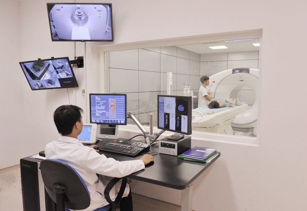
Computed tomography angiography of the lower extremities should be performed sequentially from preparation to imaging and outcome evaluation. This helps to ensure both image quality and patient safety. The procedure to perform a computerized tomography vascular scan of the lower extremities includes the following steps:Prepare equipment and tools: The computerized tomography scanner to examine the lower extremity vasculature requires a multi-slice machine with at least at least 64 ranges to accommodate high flow rates in the arteries. In addition, before the procedure, it is necessary to prepare some other medical supplies such as contrast medium, physiological saline, antiseptic alcohol, syringe, cotton, gauze, anti-shock medicine box ready for the case of Accidents occurred during and after the end of the technique.
● Prepare the patient: This is an important step that should not be overlooked in clinical practice. Patients need to be guided and consulted about the steps to be taken as well as possible complications when agreeing to have a computerized tomography scan of the lower extremities. Before entering the imaging room, the patient will be asked to remove all jewelry and personal items, change clothes in the imaging room. Instruct the patient to fast for 4 hours before performing the technique. In case the patient is too nervous and worried, some medical facilities may agree to let a family member in with the patient (wearing protective clothing) or use sedatives for the patient to cooperate. during the computed tomography scan.

Bác sĩ có thể chỉ định bệnh nhân sử dụng thuốc an thần trong trường hợp bị kích động
● Place the patient supine on the table with legs extended toward the machine chamber and head on the opposite side. Hands should be placed at head level. Establish two lines in peripheral veins, manipulation of the lower extremities should be avoided. Control the shooting table towards the camera and locate the position to capture.
● Crop the image slices horizontally and vertically with an average thickness of about 1mm each. Slices were performed without contrast first, followed by images at different phases after intravenous contrast injection.
● Evaluation of images after taking: The image after taking is a reconstructed image in three-dimensional space based on previously taken cross-sections and processing through a machine system and specialized software. A standard image should include all anatomical components of the vascular system of the lower extremities. Narrow, embolized, atherosclerotic, malformed, aneurysm lesions need to be clearly reproduced on radiographs to help diagnose many lower extremity vascular diseases.

Hình ảnh từ chụp cắt lớp vi tính hệ mạch máu chi dưới được dựng lại trên không gian ba chiều
Vinmec International General Hospital with a system of modern facilities, medical equipment and a team of experts and doctors with many years of experience in medical examination and treatment, patients can rest assured to visit. examination and treatment at the Hospital.
To register for examination and treatment at Vinmec International General Hospital, you can contact Vinmec Health System nationwide, or register online HERE.
LEARN MORE
Learn how Lower extremity computed tomography angiography digitizes the background and treats acute thrombosis of the extremities Computed tomography - the gold standard in the diagnosis of coronary artery disease


