This is an automatically translated article.
When it comes to u, we all have concerns about whether this is cancer or not? Kidney tumors, too, will include benign and malignant renal tumors. This article provides background information about a type of benign kidney tumor, renal angioma (AML).
1. What is renal lipoma?
Renal vascular lipoma (AML) was first discovered in 1911 by Fischer and in 1951 was named renal lipoma by Morgan. Renal lipomatosis is a type of benign renal tumor containing vascular, smooth muscle, and fatty components.There are two common types of AML in clinical practice:
Renal vascular lipoma with tuberous sclerosis (20%): Often with both kidneys, the clinical presentation of the disease is characteristic and easy to diagnose. Often grows large and causes bleeding. Renal lipoma without tuberous fibrosis (80%): Usually occurs in only 1 kidney, has few symptoms and is more common in women than in men. Renal lipomatosis is invasive in nature, when it grows, it will spread into the perirenal parenchyma or renal sinus, even if the later stages of the tumor can spread into nearby lymph nodes.
2. Symptoms of renal vascular lipoma
There are some cases of small renal vascular lipoma, which progressed silently and was only discovered incidentally through abdominal ultrasound.
Some other cases will have characteristic symptoms. Like other types of tumor, renal vascular lipoma will have symptoms of mass, compression. Due to its location in the kidney, there will be additional kidney-urinary symptoms associated with it. Common symptoms are: (according to Steiner's study in 1993)
Abdominal pain (80%): severe pain in the lumbar fossa region, dull pain, pain increasing gradually due to the increase in size of the tumor. If the tumor is enlarged and puts pressure on the urinary tract, it can cause renal colic. Enlarged kidney tumor (50%): Palpation of the abdomen may reveal an abnormal mass. Hematuria (25%). (5%) In case the tumor appears in one or two kidneys and grows too large, it can cause kidney failure.

Đau nhiều ở vùng hố thắt lưng là dấu hiệu của u cơ mỡ mạch thận
3. Tests in renal vascular lipoma
Imaging now plays an extremely important role in the early diagnosis of renal lipoma. Help improve disease prognosis and reasonable treatment.
Tests in renal lipoma include:
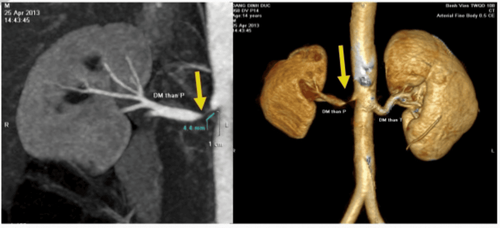
Ultrasound diagnosis of renal lipoma: This is a non-invasive method, with high sensitivity and specificity, so it is indicated first in suspected cases. renal vascular adiposity. Ultrasonography shows a very specific hyperechoic image of adipose tissue. Computed tomography (CT-Scan): Very high accuracy, up to 90%. The density of the grease is -15 to -80 Hounsfield units. Renal angiography: This is a method that both examines the blood vessels of the kidney and helps in therapeutic intervention. A large tumor can be seen in the renal parenchyma, with many malformed blood vessels inside. Kidney biopsy: Renal biopsy for histopathology is the gold standard for diagnosis. However, this method is only used when the diagnosis is in doubt and it is necessary to exclude kidney cancer.

Sinh thiết thận làm mô bệnh học là tiêu chuẩn vàng để chẩn đoán u cơ mỡ mạch thận
4. Treatment of renal vascular lipoma
Renal lipomatosis with small size < 4 cm is not indicated for treatment because it is a benign tumor and usually does not cause dangerous complications. Just follow-up at home and follow-up regularly after 6 months - 1 year to re-ultrasound to see if the tumor has increased in size or not.
With large tumors > 4 cm in size, the risk of bleeding is usually quite high and there is a risk of death if not intervened. Therefore, removing the tumor is necessary to avoid the risk of life-threatening complications such as tumor rupture, bleeding, massive hematuria, large retroperitoneal hematoma, ...
The two treatment methods for renal lipoma are total or partial nephrectomy and endovascular intervention:
Surgical removal of the tumor, total or partial nephrectomy: Current nephrectomy very limit. When the contralateral kidney is in good working order, nephrectomy is only indicated when cancer is clearly identified by immediate biopsy or when the bleeding is severe and cannot be controlled by selective embolization. Surgery has many complications during surgery such as the risk of a lot of blood loss, a large hematoma and the need to remove the entire kidney during surgery is very high. Endovascular intervention: Endovascular intervention and selective embolization are preferred because of their minimally invasive nature and ability to help patients preserve the kidney. In this case, the embolization materials used were embozene microspheres (distal occlusion) and PVA resin (proximal tumor occlusion). Detachable coils are also used to plug an aneurysm detected during angiography, minimizing the risk of future bleeding. In addition, the patient's renal function is also preserved maximally thanks to the correct selection of the tumor feeding branch and the preservation of the vascular branches of the normal renal parenchyma. Studies around the world have demonstrated the high effectiveness of endovascular intervention in hemorrhagic AML with very low rates of rebleeding and complications. Another study showed that the tumor size decreased by 33-43% after 6 months of intervention, further reinforcing the effectiveness of this method. In summary, renal lipomatosis is invasive in nature, as it grows, it will spread into the perirenal parenchyma or renal sinus, even if the later stages of the tumor can spread into nearby lymph nodes. In cases where the tumor appears in one or two kidneys and grows too large, it can cause kidney failure. Therefore, when there are warning signs of the disease, you should go to a medical facility for proper diagnosis and treatment.
At Vinmec International General Hospital, there is a package of urological examination and screening to help customers detect diseases early and have effective treatment and prevent dangerous complications. When choosing the urological examination and screening package at Vinmec, customers will receive:
Specialized urological examination; Ultrasound of the urinary system; Quantification of total PSA; Quantification of free PSA; Urine culture. To help detect possible urinary tract diseases early. Especially prostate diseases (benign prostatic hypertrophy, prostate cancer) and urinary stone pathologies.... thereby helping customers to take preventive measures.
Please dial HOTLINE for more information or register for an appointment HERE. Download MyVinmec app to make appointments faster and to manage your bookings easily.


