This is an automatically translated article.
Posterior vena cava is a rare congenital anomaly that is more common in males than in females. In normal anatomy of the urinary system, the ureter is located lateral to the vena cava, but in posterior vena cava disease the ureter passes posteriorly to and within the vena cava. This condition causes pressure on the ureters, blocking urine.
1. Normal anatomy of the urinary system
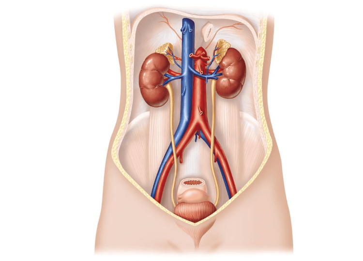
The urinary system includes the following organs: Two kidneys have the function of excreting and filtering urine, two ureters have the function of carrying urine from the kidneys to the bladder, one bladder is to store urine, one urethra is the the tube that carries urine from the bladder to the outside.
Normally, 2 ureters connect to the kidney at the position of the renal calyx, flow along the outside compared to the inferior vena cava, then cross through the iliac vessel bundle, and then connect to the bladder. The length of the ureter is about 25-28cm, the diameter is about 5mm.

Hình ảnh giải phẫu hệ tiết niệu bình thường
2. What is posterior vena cava ureteral disease?
Posterior vena cava disease is a condition in which the ureter is not in the correct anatomical position, but crosses behind the vena cava, bends around, and then goes out in front of the inferior vena cava to return to its normal path. Normally, this condition interferes with the flow of urine, causing urine to stagnate and causing hydronephrosis and dilatation of the renal pelvis.
This is a rare birth defect with a frequency of about 1 in 1000 children, caused by anomalies of the inferior vena cava during embryonic development, this phenomenon is common in the right ureter, combined with inversion of the inferior vena cava or the case of two inferior vena cava.
Based on the diagnostic method by UIV, divide posterior vena cava ureter into 2 types:
Type 1: Also known as low ring type is the common type with the rate of about 90% of cases, with pictures typical on the UIV film looks like a saxophone. In this type 1, the point of obstruction is in the ureter at the level of the 3rd dorsal vertebra. Type 2: Also known as high ring, this type is less common. With the crescent-shaped UIV image, the obstructed site is elevated, so the UIV is limited in examining the distal ureter than the CT scan.

Niệu quản sau tĩnh mạch chủ loại vòng thấp xảy ra thường xuyên hơn
Common manifestations in patients with posterior vena cava ureters include:
Dull pain in the right lumbar fossa. May be accompanied by urinary disorders such as dysuria, urinary frequency. If there is a urinary tract infection, the patient develops fever, cloudy urine, and blood in the urine. On ultrasound, there is an image of dilated renal calyces depending on the degree of obstruction, often no ureteral stones are detected. Some cases may have complications and stone formation, so it is easy to misdiagnose. In cases with these birth defects, the sooner the treatment is to re-establish the circulation of urine, the better, to avoid long-term complications and affect kidney function.
3. Methods of diagnosis of posterior vena cava ureteral disease
In addition to relying on clinical manifestations, the use of laboratory measures is essential to be able to make a definite diagnosis. When there are clinical signs and suspicious ultrasound images, the doctor will order the following paraclinical tests:
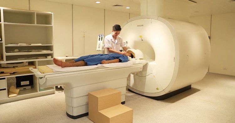
UIV (Intravenous urogram): On the film, there are saxophone signs or imaging signs. the hook. CT scan: is the preferred diagnostic tool because in addition to determining the disease state, the path of the ureter also evaluates the status of the inferior vena cava, the relationship between the inferior vena cava and the ureter, Also evaluate the organs and organs around the ureter. MRI: Has diagnostic value equivalent to CT scan.

Chẩn đoán bệnh niệu quản sau tĩnh mạch chủ bằng hình ảnh
4. Methods of treatment for posterior vena cava disease
For patients with ureters behind the inferior vena cava, the indicated treatment is surgery.
The purpose of surgical treatment of the disease is to return the ureter to its normal position and reestablish the urethral circulation, avoiding urine stagnation in the kidney.
The surgical methods used for treatment include:
Open surgery: Open surgery to reconnect the ureter, bringing the ureter to the correct position. However, this method has some disadvantages such as large incisions, slow postoperative recovery, and increased hospital stay. Laparoscopic or retroperitoneal surgery: Reposition and anastomosis of the ureter to treat the disease. Compared with open surgery, laparoscopic surgery has more advantages such as less invasive method, small incision, high aesthetics, faster recovery time. Robotic surgery: There are a number of hospitals that have used robots to conduct surgery, also with good results. Posterior vena cava ureteropathy is a rare congenital anomaly that often causes renal stasis. There are some cases where the disease is difficult to detect, so regular health checks are also a method of early detection of the disease before complications occur. The disease is treated by surgical methods, with high treatment efficiency.
Vinmec International General Hospital with a system of modern facilities, medical equipment and a team of experts and doctors with many years of experience in neurological examination and treatment, patients can completely peace of mind for examination and treatment at the Hospital.
To register for examination and treatment at Vinmec International General Hospital, you can contact Vinmec Health System nationwide, or register online HERE.
Recommended video
Fetal screening - A healthy baby is born
MORE
Learn about vesicoureteral reflux in children What is an ectopic ureter? Diagnosis and treatment of vesicoureteral reflux


