This is an automatically translated article.
Intracranial hypertension syndrome can cause cerebral edema, cerebral ischemia, or very rapid brain collapse that can cause death or irreversible damage, so it should be diagnosed early with diagnostic tests for the syndrome. increase intracranial pressure and manage aggressively.
1. Causes of raised intracranial pressure syndrome
Normally, the intracranial pressure measured in the ventricles ranges from 10 to 15 mmHg (136 to 240 mm H2O). Elevated intracranial pressure is defined as persistent elevation of pressure above 20 mmHg/cmH2O
Causes of ICP syndrome include:
Traumatic brain injury Brain bleeding: In brain parenchyma, ventricles , subarachnoid bleeding Large branch occlusion of cerebral artery: Occlusion of internal carotid artery, middle cerebral artery... Brain tumor Nervous infection: Encephalitis, meningitis, brain abscess Hydrocephalus.

Viêm màng não là một trong những nguyên nhân gây ra hội chứng tăng áp lực nội sọ
Other likely causes of increased intracranial pressure:
Hypercapnia; hypoxemia Ventilation using high PEEP (positive end-expiratory pressure) Hyperthermia Hyponatremia Convulsive status.
2. Diagnosis of raised intracranial pressure syndrome
Diagnostic tests for raised intracranial pressure syndrome may include:
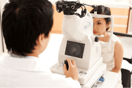
2.1 Ophthalmoscopy Depending on the degree of increased intracranial pressure, the optic nerve changes in different stages from mild to severe.
Early stage: The papilledema stage presents with the optic disc filling up to the surface of the retina and being pinker than normal. The papilledema margin fades from the nose to the temple, losing central luminosity of the congested blood vessels. Stage of papilledema: The optic shoulder is completely erased, the optic disc is swollen and swollen on the retinal surface, like a mushroom, this convexity is measured by a diopter (1mm = 3diop) of the red-pink optic disc that flares out like a flame. . The blood vessels constricted. Hemorrhagic stage: In addition to the above image, there are also hemorrhages in the optic disc and retina.

Xét nghiệm chẩn đoán hội chứng tăng áp lực nội sọ bằng soi đáy mắt
Stage of atrophy: The final stage, also known as the decompensation phase. The spines become white and silver, lose their shine, and have rough edges. The blood vessels are sparse and pale, accompanied by clinically reduced visual acuity. The papilledema in ICP syndrome is often bilateral, with varying degrees of severity. Unilateral papilledema is rare. In prefrontal lobe brain tumors, papilledema on the side of the tumor and papilledema on the contralateral side (Foster Kennedy syndrome) may be seen.
2.2 Cranial cephalometric scan Computerized tomography (CT-scan) of the skull can show:
Cerebral edema, brain structure pushed, midline structure changed. Dilated ventricles: Due to obstruction of the circulation of cerebrospinal fluid. Brain bleeding, cerebral ischemia, brain tumor, brain abscess... Cranial magnetic resonance (MRI) brain: For more information about brain damage.
Angiography when focal signs are suspected (occupation), in cases of severe cranial hypertension, the arteries lose their softness (loss of flexure). Identify cerebral vascular malformations.
2.3 EEG Nonspecific, but suggestive of localization (early stage) and assessment of severity of raised intracranial pressure, more or less slow wave, possibly bilateral, especially late stage .

Điện não đồ có thể gợi ý khu trú và đánh giá tình trạng của tăng áp lực nội sọ
2.4 Blood tests Blood tests can help detect hypernatremia syndrome due to hyponatremia.
2.5 Transcranial Doppler Blood flow rates above 150mm/s indicate vasoconstriction.
Test of cerebrospinal fluid is contraindicated in raised intracranial pressure, when more than two diopters only unless meningitis is suspected, then need to be punctured in the supine position, fine needle, drained, and taken a small amount of fluid for testing. experience.
Syndrome of increased intracranial pressure is an extremely dangerous syndrome that needs to be diagnosed quickly and promptly treated to avoid life-threatening as well as permanent brain damage.
Any questions that need to be answered by a specialist doctor as well as customers wishing to be examined and treated at Vinmec International General Hospital, you can contact Vinmec Health System nationwide or register online HERE.
SEE MORE
Learn about raised intracranial pressure syndrome What is intracranial pressure and what is normal? Signs of increased intracranial pressure


