This is an automatically translated article.
The article was professionally consulted by Specialist Doctor II Khong Tien Dat, Doctor of Radiology - Department of Diagnostic Imaging - Vinmec Ha Long International General Hospital. The doctor has more than 14 years of experience in the field of diagnostic imaging.Ultrasound is a method to help accurately diagnose many diseases. In which, renal ultrasound is used to diagnose pathologies in the urinary system. Drinking water before a kidney ultrasound for a certain period of time helps to stretch the bladder and thereby make it easier to investigate and detect diseases.
1. What is renal ultrasound?
Ultrasound is a non-invasive, painless and convenient method, cheap but has high diagnostic value. It can be performed repeatedly for diagnosis, monitoring, and combined treatment procedures. So far, there have been basically no unwanted effects of ultrasound on humans. Because of these advantages, ultrasound has been widely used as a routine test for cancer screening. and investigate many diseases in the abdomen.
Usually, when doing a general abdominal ultrasound to examine areas in the abdomen such as the liver, spleen, pancreas, etc., the doctor also examines the organs of the urinary system.
2. Kidney ultrasound technique
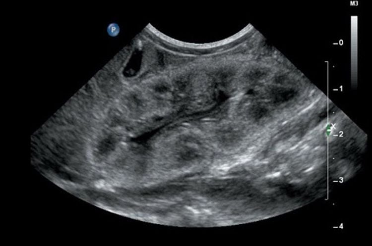
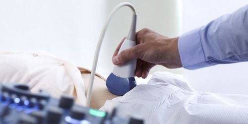
Renal ultrasound is a method of examining the urinary system with sound waves, when sound waves are sent through the transducer into the abdominal organs, they will collect images at the image processor, from those images to make diagnoses. diagnose the disease if there is any abnormality.
Renal examination techniques in renal ultrasound are as follows:
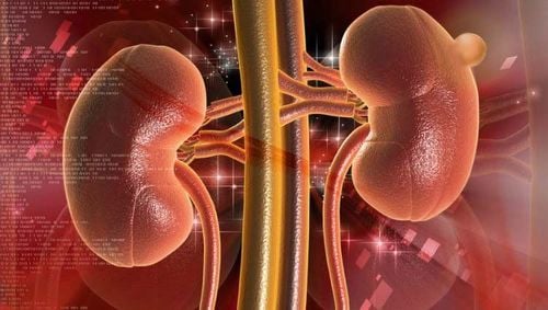
The kidneys are located in the lumbar fossa, on a horizontal plane with an oblique direction from back to front and from outside to inside. When renal ultrasound, it is necessary to evaluate the longitudinal and transverse sections of the kidney to assess the function of the kidney. When doing an ultrasound of the right kidney, the doctor usually relies on the liver window to clearly see the upper pole of the kidney and through the subcostal slice to see the lower part of the kidney. The left kidney usually has to be resected in the posterior lateral direction of the abdominal wall, because of the difficulty in using the splenic window. The ureter is the connecting structure between the kidney and the bladder, ultrasound often shows the lower renal pelvis and the proximal bladder. However, it is often difficult to observe and evaluate unless abnormalities are present. In addition, evaluate the patient's bladder.
3. What diseases can be detected by renal ultrasound?
Renal ultrasound can detect urinary tract diseases including:
Congenital structural changes in the urinary system such as: Congenital kidney disease (Double kidney, horseshoe kidney, prolapsed kidney, ectopic kidney) , renal aplasia..), ureteral pathology (ureteral prolapse, double ureter...), Bladder (double bladder, bladder diverticulum, bladder prolapse...). Renal cyst pathology: Simple kidney cyst, acquired kidney cyst, polycystic kidney... Renal tumor pathology: Benign tumor (angioma, adenoma...) and malignant tumor (cancer) renal cell carcinoma, lymphoma, etc.). Bladder cancer, an abnormal tumor in the bladder. Dilatation and occlusion of the renal calyx system: Dilatation of the renal pelvis, pyelonephritis, renal water retention... Kidney stones, ureteral stones and bladder stones. Infectious and inflammatory diseases of the urinary system: pyelonephritis, renal abscess, renal tuberculosis...

Diffuse pathology: Acute tubular necrosis, glomerulonephritis ... Urinary system trauma. Renal Doppler: Detects renal vascular abnormalities such as renal artery stenosis.
4. Kidney ultrasound should drink water?
For kidney ultrasound, the patient needs to drink enough water before the ultrasound about 1 hour to let the bladder stretch urine to help doctors easily observe the image of the kidney as well as some other organs in the urinary system. . Specifically:
When the urine in the bladder is stretched, it will create a good sound transmission environment, helping to enhance the sound for the ultrasonic waves to pass through (by providing a good environment for sound conduction). When the bladder is distended, urine will help to evaluate the internal structure of the bladder, abnormalities of the bladder. When straining the urine, using the acoustic window from the bladder to detect stones located in the distal ureter, near the bladder helps to diagnose the location and size of ureteral stones. In addition, when there is a lot of urine in the bladder, it causes the bladder to stretch, leading to a change in the position of the uterus, from which the uterus is pushed up to be easier to see. A distended bladder is easy to observe organs in the pelvic region such as the ureters, prostate, uterus, and less than 3 months of pregnancy.

5. Notes on how to drink water during kidney ultrasound
Before performing renal ultrasound, the patient should pay attention to how to drink water as follows:
Should hold urine so that when feeling urge to urinate, start the ultrasound, should not hold urine excessively, this will make the bladder Excessive stretching affects image quality. The patient feels discomfort when pressing on the bladder area for ultrasound. When the bladder is overstretched with urine, it can lead to physiological pyelonephritis. However, you do not need to worry because this condition will go away after urinating. It is recommended to drink 4-6 glasses of water about 1 hour before the ultrasound, so it will help the bladder stretch before the exam. Drinking enough water to stretch the bladder helps the ultrasound doctor to better survey the pelvic organs and give more accurate results. When there is no urine, the examination will become more difficult and it is not possible to accurately diagnose the disease, the patient needs to wait more time for the bladder to empty. Patients should actively drink enough water before the examination to get the best results and avoid having to wait for a long time during the examination.
Currently, Vinmec International General Hospital has been using modern generations of color ultrasound machines. One of them is GE Healthcarecar's Logig E9 ultrasound machine with full options, HD resolution probes for clear images, accurate assessment of lesions. In addition, a team of experienced doctors and nurses will greatly assist in diagnosing and early detection of abnormal signs of the kidney as well as of many other organs in the body in order to come up with a method. timely treatment.
Please dial HOTLINE for more information or register for an appointment HERE. Download MyVinmec app to make appointments faster and to manage your bookings easily.


