This is an automatically translated article.
The article is professionally consulted by Master, Doctor Vu Huy Hoang - Radiologist - Department of Diagnostic Imaging and Nuclear Medicine - Vinmec Times City International Hospital
The detection of diseases related to the biliary tract has become easier thanks to the advancement of science. And the method of x-ray through the bile duct through kehr is evaluated as a method of high value in diagnosis and treatment after surgery for biliary drainage. Need an x-ray after removal of the biliary tract?
Is it necessary to take x-rays after removing the biliary tract? Let's explore this question right here.
1. Principle of the Kehr . ductal X-ray method
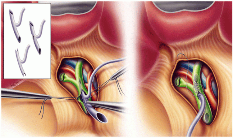
Injecting a contrast agent containing water-soluble iodine into the biliary tract, this is done under a dark screen and using a Kehr tube (postoperative). From there to also determine the extent and location of some pathologies including:
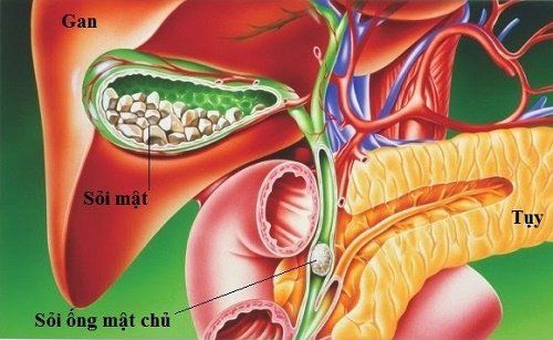
Bile leakage or biliary fistula gallstones, cholangitis, biliary tract tumors, blood clots Assess the degree of narrowing or dilatation of the intrahepatic and extrahepatic bile ducts Investigation of the circulation of bile through the sphincter of Oddi to the duodenum
2. Before taking X-ray of the biliary tree, what do you need to prepare?
Performer:
Specialist doctor Electro-optical technician Means:
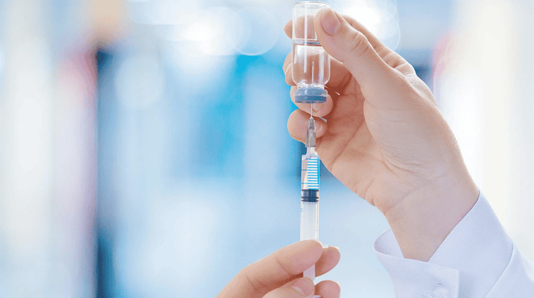
Iodine contrast agent dissolved in water Distilled water or physiological saline Syringe 20ml, needle 18-20G Gloves, mask, Caps Clamps, Bean trays Antiseptic alcohol, Cotton, Contrast first aid equipment, medicine boxes Specialized X-ray machines Films, cassettes, storage systems Consumables Patients:
Before the scan, the patient can fast or eat a light meal to avoid fermenting and fibrous food. You can view the surgical report so that you can orient the imaging position. Check the patient's information such as full name, age, address, ..., pay attention to learn more history of allergies, especially allergy to contrast drugs or drugs containing iodine. The doctor explained the imaging process and possible complications to reassure the patient.

3. Conduct X-ray of bile ducts through Kehr . duct
Preparation:
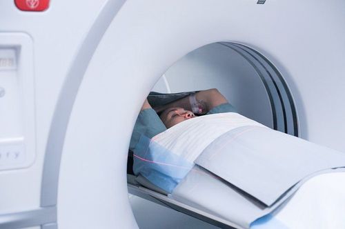
The patient will lie supine on the X-ray table, arms raised above the head, legs stretched, and the drainage bag hanging close to the table. The doctor wears gloves, a hat and a mask. Take about 5mL of iodinated contrast agent with a concentration of 300-400 mg/mL, mix it with 0.9% NaCl solution in a ratio of 1⁄3 or 1⁄4. Pay attention to dilution to limit contrast agents to obscure the gallstone tract. Or can use contrast agent with concentration of 120mg/mL and mixed with physiological saline. Prepare the Kehr drain:
Pay attention to stroke the Kehr tube so that the bile can flow out, expel the gas, check carefully in the tube until there are no air bubbles. Clamp the Kehr tube about 3 to 5cm away from the patient's skin to avoid backflow as well as reduce the amount of drug residue in the tube. Use an iodized alcohol solution to disinfect the upper part of the clamp. Then proceed to inject 20ml of iodine contrast solution slowly into the Kehr tube through the sterilization site. (Note: the syringe must be raised at an angle of more than 45 degrees to prevent gas from entering the biliary tract). In order to detect and promptly handle cases of allergies or complications with contrast agents, patients must be monitored during the implementation process. Perform cholangiography through Kehr's duct:
Adjust the patient's position so that the patient lies on his left side so that the contrast agent can easily enter the left hepatic bile duct, then lie on his back for the scan. At this time, the doctor monitors on the screen, if the drug fills the entire biliary tract, ask the patient to hold his breath. The purpose of imaging and preliminary diagnosis is to choose the appropriate positions to expose the lesions in the biliary tract.
Shooting positions
Straight position for overview survey of the entire bile duct Right oblique position posteriorly for the purpose of surveying the right hepatic bile ducts Left oblique position posteriorly to examine the left hepatic bile ducts Straight position to observe the drug circulation down to the duodenal area Right slant position to easily survey the location of biliary tract injury After taking X-ray, proceed to smoke the contrast in the biliary tract, Disinfect, remove clamp.
Results of radiographs
Must adjust the appropriate contrast The film i shows the entire biliary tract and the outside of the liver. It can be said that the method of X-ray cholangiography through the Kehr duct is intended to help patients early detect diseases related to the lost sugar. With the information shared above, hopefully you have found yourself the answer to whether you need an X-ray after removing the bile duct or not. Currently, X-ray is a diagnostic imaging method performed routinely at Vinmec International General Hospital, including X-ray technique after removing the biliary tract. The X-ray procedure at Vinmec is carried out methodically under the guidance of the medical team of the Department of Diagnostic Imaging. In addition, Vinmec is now equipped with the most modern X-ray machine, meeting international standards for true and clear images, helping doctors to accurately diagnose the disease and the stage of the disease, from It has an effective treatment direction, creating a sense of safety for the patient.
Any questions that need to be answered by a specialist doctor as well as customers wishing to be examined and treated at Vinmec International General Hospital, please register for an online examination on the Website for the best service.
Please dial HOTLINE for more information or register for an appointment HERE. Download MyVinmec app to make appointments faster and to manage your bookings easily.


