This is an automatically translated article.
The article was professionally consulted by Specialist Doctor I Tran Cong Trinh - Radiologist - Radiology Department - Vinmec Central Park International General Hospital. The doctor has many years of experience in the field of diagnostic imaging.If not diagnosed and detected in time, diseases related to circulation and veins easily cause many dangerous complications, difficult to treat. One of the modern techniques to help quickly detect abnormalities in the circulation is Doppler ultrasound of blood vessels.
1. Overview of vascular ultrasound
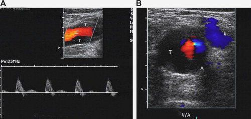
According to the principle of the Doppler effect, when a beam of ultrasonic waves is transmitted to an object, there will be an acoustic reflection phenomenon. The frequency of the reflected ultrasonic beam will change compared to that of the transmitted beam if the relative distance between the source and the object changes, the frequency increases if the distance decreases and vice versa.Doppler ultrasound of blood vessels is a method of using sound waves to examine and evaluate the body's circulatory system. The ultrasound process helps to simulate the image, structure, size and circulation of arteries and veins to identify blockages and detect the location of blood clots, if any.
Doppler ultrasound techniques of blood vessels:
Continuous Doppler ultrasound: Continuous Doppler with a probe with 2 crystals, emits and receives continuous feedback waves to help measure very large blood flow velocities. However, it cannot record the speed at a certain point, but only records the average speed of many moving points that the emitted sound wave encounters along the way. Pulsed Doppler ultrasound: With a 1-crystal transducer that both has the function of transmitting and receiving feedback waves. Sound waves are delivered in pulses along the probe scan direction, but only the echoes from the window placement are recorded and processed. The received pulsed Doppler signal is presented in audio, spectral and color form. Different Doppler frequencies are caused by red blood cells moving at different velocities, so the Doppler spectrum helps to recognize the velocity of the flow. Color Doppler Ultrasound: Is a color-coded pulsed Doppler image overlaid on a 2-D ultrasound image. Power Doppler ultrasound: Only examines the level of the Doppler signal without regard to the direction of flow. More sensitive than color Doppler for flow detection, but susceptible to motion artifacts.
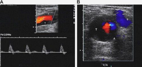
Siêu âm Doppler mạch máu là phương pháp sử dụng sóng âm để kiểm tra và đánh giá hệ thống tuần hoàn của cơ thể
2. When is vascular ultrasound needed?
Doppler ultrasound of blood vessels is a very valuable non-bleeding method, not only to help evaluate vascular lesions in terms of anatomical morphology, but also to help check the flow of blood in tissues and organs. From there, detect abnormalities such as blockage of arteries, veins, atherosclerosis, blood clots... Vascular ultrasound not only assesses the level and risk of cardiovascular diseases but also helps evaluate the effectiveness of previous treatments.Specifically, vascular ultrasound is often done to:
Assess the state of blood circulation to organs and tissues in the body. Detect and locate blockages and abnormal plaques and support treatment plans. Detects blood clots in blood vessels. Assess the ability to perform coronary angioplasty of patients with related diseases. Evaluation of the effectiveness of vascular transplantation or vascular bypass surgery. Detection of aneurysms, arteriosclerosis. Detection and assessment of varicose veins, deep vein insufficiency, deep vein thrombosis. The requirement is that the doctor performing vascular ultrasound needs to master the principles and techniques of ultrasound and must have a solid knowledge of circulatory and vascular pathology.
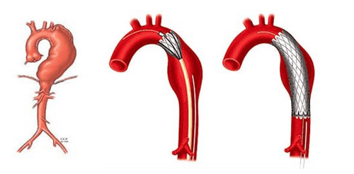
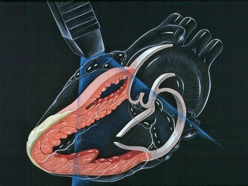
Siêu âm mạch máu thường được thực hiện bệnh lý phình động mạch
3. Prepare in advance for vascular ultrasound
Prepare patient:Wear loose, comfortable clothing, remove jewelry to facilitate the ultrasound. Be introduced to the process and purpose of vascular ultrasound. Limit continuous movement during ultrasound. Technician performing:
1 specialist trained in vascular ultrasound. Means of use:
Ultrasonic probes of all kinds: 1 flat probe (frequency 7.5-12.5 MHz), 1 fan probe (frequency 3.5-5 MHz), pencil probe , special probe (if needed). Doppler ultrasound machine with full pulse ultrasound modes, color ultrasound, energy ultrasound.
4. Vascular ultrasound procedure
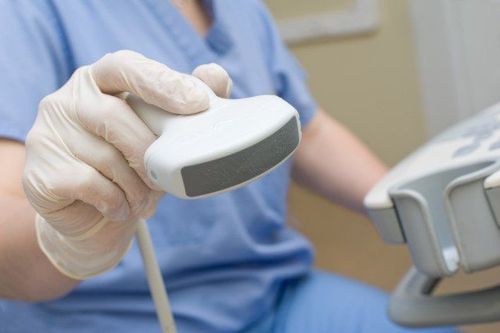
The procedure of vascular ultrasound is very simple:The patient will be instructed to lie on his or her side or face down on a flat surface. After that, the patient is applied a water gel to the areas that need ultrasound, the doctor will move the transducer in this area to obtain the necessary imaging images. Once the ultrasound has been completed, the patient changes clothes and waits for the doctor to review the resulting images. The entire vascular ultrasound process usually takes 15-45 minutes.
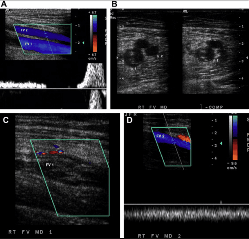
Quy trình siêu âm mạch máu diễn ra rất đơn giản
5. Is vascular ultrasound painful?
Vascular ultrasound is a test technique that does not use ionizing radiation, which helps to provide detailed images of soft tissues that cannot even be shown on an X-ray. Doppler ultrasound of blood vessels does not cause pain and does not cause any adverse effects on the patient's health.Vinmec International General Hospital with a system of modern facilities, medical equipment and a team of experts and doctors with many years of experience in medical examination and treatment, patients can rest assured to visit. examination and treatment at the Hospital.
Please dial HOTLINE for more information or register for an appointment HERE. Download MyVinmec app to make appointments faster and to manage your bookings easily.


