This is an automatically translated article.
The article is professionally consulted by Master, Doctor Pham Manh Chung - Department of Diagnostic Imaging - Vinmec Ha Long International HospitalRenal function scintigraphy is a simple diagnostic technique, easy to conduct, very valuable in kidney diseases. Renal scintigraphy not only provides information about the function of individual kidneys through quantitative and qualitative analysis, but also provides information about the location, size and anatomy of the kidney. In addition, this test is very useful in cases where the patient is sensitive to high-iodine or urea-based contrast and cannot be radiographed.
1. What is a kidney scan?
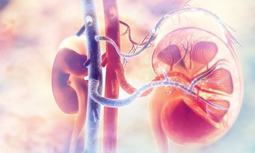
Scanning is a method of imaging the specific distribution of radioactive substances inside organs by measuring their radioactivity from outside the body. Scanning is not only a simple imaging method of morphology, but it also helps to evaluate the function of the organ and a number of other pathological changes of the organ itself.
Renal function scintigraphy, including mass renal imaging and isotope nephrography, is currently widely used in nuclear medicine departments. With the help of modern high-precision measuring machines and the use of radioactive tracers for high-quality images, renal scintigraphy has become an indispensable technique in the complex of medical examinations. kidney function test, contributing to improving the efficiency in the diagnosis of kidney and urinary tract diseases.
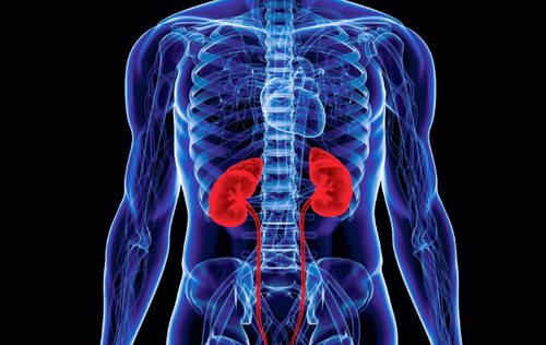
The principle of renal function scanning technique is to use substances whose only route of excretion from the body is through the kidneys, radiolabeled isotopes, and then intravenously administered to the patient. Record kinetic images and graphs of radioactivity over time of each kidney, qualitative and quantitative analysis, thereby evaluating the function of each kidney individually.
2. Indications for kidney scintigraphy

Bệnh nhân đang có thai không được chụp xạ hình thận vì sẽ dùng dược chất phóng xạ.
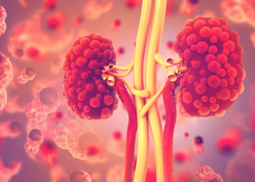
Assess the function of both kidneys or each kidney separately. Evaluation and monitoring of kidney diseases such as renal hypertension, pyelonephritis, glomerulonephritis, tubular necrosis, acute renal failure... Evaluate and monitor urinary tract obstruction. Suspicious on one side. Kidney injury. Evaluation and follow-up after kidney transplantation. Renal scintigraphy is contraindicated in the following cases:
The patient is taking ACE inhibitors. Patient is pregnant or lactating.
3. Kidney X-ray procedure

Bệnh nhân được tiêm tĩnh mạch đồng vị phóng xạ, ghi lại hình ảnh động học và đồ thị hoạt độ phóng xạ theo thời gian của từng thận, phân tích đồ thị và hình ảnh, qua đó sẽ đánh giá được chức năng của riêng từng thận.
Need to remove jewelry that could interfere with the scan, may have to take off all or most of the clothing, depending on what area is being examined (underwear may be allowed if it doesn't interfere with the scan). interference with scans).
The technician cleans where the radioactive tracer will be injected on the arm. A small amount of radioactive tracer will be injected, and a diuretic may also be injected. The patient can lie supine on the table, standing or sitting upright. A large scanning camera is placed close above the abdomen and scans for radiation immediately after the radioactive tracer is injected. Scans can be performed every few minutes for about 30 minutes.
Multiple images can be taken 1 to 2 hours after the tracer is injected. The scans create images as the tracker moves through the kidneys. A histogram can be made using the information from the kidney scan by plotting the tracer movement through the kidney and recording it on the chart. These records provide information about different stages of blood flow and kidney function.
A kidney scan usually takes about 30 minutes to 1 hour. It is necessary to stay still during each scan to avoid blurring the image. The camera does not produce any radiation, so there is no further exposure to radiation while the scan is being performed.
4. Is kidney scan dangerous?
The patient may feel nothing from the needle puncture when the tracer is injected or feel a brief sting when the needle is inserted through the skin. The kidney scan is usually painless. Sometimes the patient may find it difficult to stay still during the scan, adding pillows or blankets that make them most comfortable before the scan begins. Try to relax by breathing slowly and deeply.
Allergic reactions to radioactive tracer are very rare. Most tracer will be eliminated from the body through urine or feces within 1 day. Therefore, it is important to wash the toilet immediately and wash your hands thoroughly with soap and water. The amount of radiation is so small that there is no risk to anyone coming into contact with the patient after the test.
Sometimes soreness or swelling may occur at the injection site. These symptoms are usually relieved by applying a warm, moist compress to the arm.
There is always a very small risk to cells or tissues from being exposed to any radiation, including low levels of radiation due to the radioactive tracer used for the scan.
Kidney imaging technique is being applied by Vinmec International General Hospital in the diagnosis of urinary tract infections and kidney failure. Vinmec uses the most modern SPECT/CT Discovery NM/CT 670 Pro system with the most modern 16-series CT in Southeast Asia, manufactured by the world's leading medical equipment company GE Healthcare (USA). The team of doctors and technicians at Vinmec are experienced, well-trained at home and abroad, bringing maximum efficiency to the imaging process.
Please dial HOTLINE for more information or register for an appointment HERE. Download MyVinmec app to make appointments faster and to manage your bookings easily.