This is an automatically translated article.
This article is professionally consulted by Dr. Pham Quoc Thanh - Radiologist - Department of Diagnostic Imaging - Vinmec Hai Phong International General Hospital.1. Benefits of venous magnetic resonance imaging
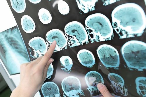
Magnetic resonance imaging, also known as MRI for short, is a modern image processing technique and is considered a great revolution in medicine. To date, MRI is increasingly being used in countries around the world because of its very high safety and accuracy, along with its non-invasive nature because it does not use X-rays. Magnetic resonance imaging (MRI) always gives high resolution and can survey multi-sections, providing the sharpest and most realistic images of the location to be taken, and the successful assessment of the properties of the required tissue. must survey. From there, it brings beneficial value to the diagnosis and treatment process of the specialist about the current condition of the veins.The great benefits that magnetic resonance equipment brings include:
Patients will not have to suffer health effects due to radiation in X-ray and CT scan methods. The patient is not subject to biological effects. Get quality, multi-plane images like vertical plane, horizontal plane, forehead plane and any other inclined plane. The resolution of soft tissue is relatively high. The displayed image quality is better than conventional shooting methods. Cerebral angiography (MRA) is possible, even without the use of additional contrast agents. This is a non-invasive imaging technique that does not cause pain and unwanted side effects to the patient. Contrast agents have very rare side effects.
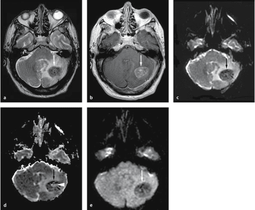
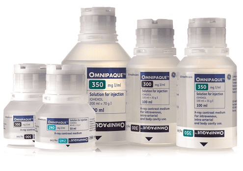
Magnetic resonance venography has Magnetic contrast injection is this non-invasive imaging test that will allow detailed examination of the entire venous system, capable of detecting many pathologies such as venous malformations, arteriovenous malformations or evaluating venous thrombosis. With contrast, contrast agents can help differentiate or "enhanced contrast" the vein from the surrounding tissue.
Therefore, you should contact medical centers in advance before scheduling an examination, taking an MRI for detailed advice and support about contraindications in this technique.
2. What patients need to prepare before performing an intravenous MRI
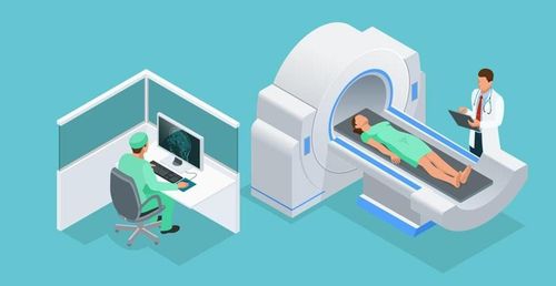
During the MRI scan, the patient lies inside a very large magnet tube, where there is a very strong magnetic field. Therefore, you need to follow the instructions of the radiologist and doctor.During the scan, the patient needs to lie on his back, not moving, to get the best quality images with the best effect.
Patients should bring health records such as laboratory results, ultrasound, CT film,... so that specialists can refer to and make decisions about appropriate imaging techniques. best. After completing the procedure, you will change into the clothes of the MRI room and remove the metal belongings, electronic devices such as watches, phones, headphones, ... In the MRI room, there will be installations Specialized tools for checking foreign bodies or small metal devices in the body in organs with loose tissue such as the heart, lungs, eyes, brain, ... these cases should not be performed. Magnetic Resonance. For cases requiring contrast injection, staff will check the patient's history of drug allergies, along with possible drug allergies. Note that the contrast agent is non-toxic and does not affect the health of the user.
3. Intravenous magnetic resonance imaging procedure with magnetic contrast injection
The patient was instructed to lie on his back for an intravenous line with an 18G needle.Then fitted coils that attract signals to all body positions.
In addition, the patient can wear noise-cancelling headphones if desired.
Take pulses localized in the area to be examined (eg lower limb, upper limb) with three planes. Capture the venous pulse according to the pulse receiver at the periphery. Capture venous pulses when contrast is injected. Contribute to the reproduction of multi-faceted together with three-dimensional space. Evaluation of the results obtained:
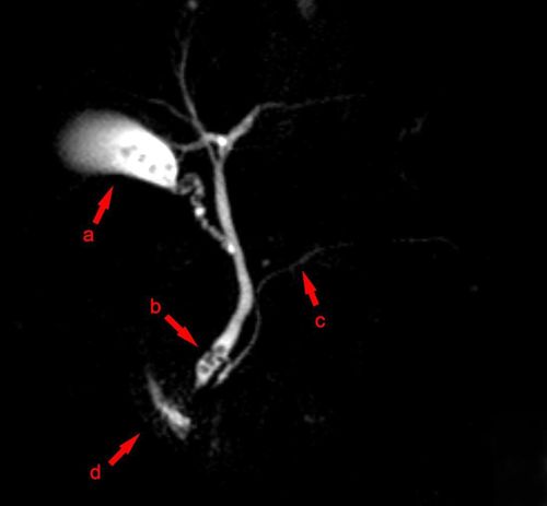
Detect lesions in the veins if any. The image will clearly show all the anatomical structures at the vein site being examined. The specialist doctor will rely on the sharp images obtained from the magnetic resonance imaging machine to describe on the internal computer and print the results to advise the current health status of his patients. Management of complications in MR imaging:
If the patient is scared or agitated, give encouragement and comfort to help them regain their composure. Complications directly related to the use of contrast agents: it is necessary to strictly follow the direction of the specialist, according to the standard procedure in the diagnosis and management of adverse events from contrast agents. It can be seen that this is one of the important techniques in diagnostic imaging to accurately assess the lesions and abnormalities of the venous system, which can replace ultrasound, computed tomography and digitization. erase background. The scan time is about 45-60 minutes and the patient will receive the MRI results after 15-30 minutes, depending on the abnormalities in the body of the scanner.
The MRI scanner will evaluate the entire venous system in a specific, multi-plane, 3D rendering. Drugs used in the examination of the venous system will not cause complications and affect the patient's health.
Vinmec International General Hospital is a unit that has a 3.0 Tesla magnetic resonance imaging machine system with modern Silent technology that brings outstanding advantages and owns an imaging room that meets the standards of the Ministry of Health. Above is the procedure for intravenous magnetic resonance imaging with injection of magnetic contrast, along with the notes that the patient needs to prepare before conducting this advanced technique. Currently, Vinmec International General Hospital has installed a 3.0 Tesla magnetic resonance imaging machine with the most modern Silent technology in our country, providing the best quality. Therefore, customers can be assured of the results of examination and treatment when performed at Vinmec.
Master. Doctor. Pham Quoc Thanh has received intensive training and participated in many national and international scientific conferences on diagnostic imaging. The doctor has 13 years of experience in the field of diagnostic imaging and is currently a doctor at the Department of Diagnostic Imaging, Vinmec Hai Phong International General Hospital.
Please dial HOTLINE for more information or register for an appointment HERE. Download MyVinmec app to make appointments faster and to manage your bookings easily.


