This is an automatically translated article.
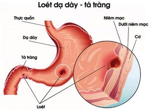
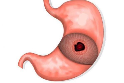
Perforation of duodenal ulcer is an acute complication of peptic ulcer disease. The disease needs to be treated promptly to minimize the danger. Patients with perforated duodenal ulcer will be treated to suture the perforation, surgically manage perforation complications and treat ulcers.1. When is the indication for endoscopic treatment of perforated duodenal ulcer?
Endoscopic treatment of perforated duodenal ulcer is indicated for patients with a diagnosis of perforation of duodenal ulcer alone. Some patients need contraindications to this treatment method, including:The patient's condition is too weak, there are too many co-morbidities The patient has a history of surgery for peritonitis, intestinal obstruction Patients with ascites Free or localized ascites Patients with abdominal wall hernia, umbilical hernia Infection localized abdominal wall Have coagulopathy Patients with coronary artery disease, valvular heart disease, chronic heart disease contraindication to intra-abdominal ventilation Before endoscopic treatment of perforated duodenal ulcer, patients need to perform basic tests as prescribed by a gastroenterologist, endoscopy and anesthesiologist to ensure they are eligible to perform. Endoscopic. In addition, patients need to take X-ray of the abdomen without preparation, use prophylactic antibiotics before surgery.
2. Conduct endoscopic treatment of perforated duodenal ulcer
Nội soi điều trị thủng ổ loét hành tá tràng được chỉ định cho người bệnh có chẩn đoán thủng ổ loét hành tá tràng đơn thuần
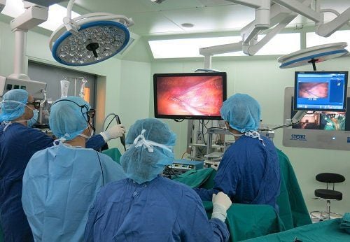
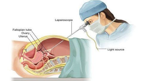
Perform Hasson type open air inflating with pressure of 12mm Hg, flow rate of 2.5 l/h. Through an incision below the umbilicus or above the umbilicus (the length of the incision depends on the diameter of the trocar intended to be used) use a Veress needle to inject CO2 into the peritoneal cavity with a pressure of 9 to 12 mmHg). The Hasson Canule is an alternative to the Verres needle. Usually 4 trocars, 3 10mm trocars and 1 5mm trocar are used.
The first 10mm trocar is used for the bronchoscope placed at the umbilicus, the 10mm bronchoscope inclined at 30 degrees is used. After placing the first trocar, performing a full abdominal view, laparoscopy in itself is a valuable diagnostic tool, so a thorough examination of the entire abdomen is necessary. Other trocars were placed under endoscopic guidance, including a 10mm epigastric trocar that used a hepatic retractor and round ligament to expose the anterior aspect of the stomach, pylorus, and duodenal bulb.
Evaluation of perforation status includes location, diameter, and degree of fibrosis affecting surrounding organs, with gastric perforation to evaluate for malignancy. Assess abdominal status, degree of dirt, pseudomembranous.
Trocar 10mm for manipulation placed above the left breast line, 5cm below the costal margin. The assistant trocar is placed in the right anterior axillary line at the level of the navel.
Stage I: Absorb food, dirt, and pseudomembranous membranes in the abdomen. Stage II: With gastric perforation, it is necessary to trim the perforation margin for a systemic biopsy. Enhanced suture with omental patch, 3 stitches through the duodenum visible on both sides of the perforation and tightened to close the perforation. Endoscopic suture of the omental stalk over the perforation completes closure. When the omentum is small, the crescent ligament can be used instead.
Perforation stitching method: Using Vicryl 2.0 or 3.0 slow-absorbing metal thread about 18 - 20cm long is enough. Using the liver lift to lift the underside of the liver to reveal the perforation, the perforation was sutured. If the perforation is <0.5 cm, suture an X, the direction of suture follows the axis of the digestive tract, then tighten the suture to close the hole.
Stage III: Abdominal lavage The peritoneal cavity is one of the most important parts of surgery, accounting for a significant part of the surgery. Wash to ensure all dirt and fascia in the abdomen. Wash each cavity in the abdomen in combination with changing the patient's position to clean the cavity. Particular attention should be paid to the washing of the suprahepatic and subhepatic spaces, the lateral sulcus, the left subdiaphragmatic space, and the right iliac fossa. If the abdominal cavity is clean, there is no need for drainage; if there is late peritonitis, the right subhepatic drainage should be placed. The peritoneal cavity is usually drained. On the other hand, some authors do not advocate drainage of the peritoneal cavity.
IV phase: Withdraw the trocar, deflate and close the holes
3. Monitor and manage complications after laparoscopic treatment of perforated duodenal ulcer

Sau khi mổ nội soi bệnh nhân sẽ được sử dụng thuốc kháng sinh trong vòng 5 ngày hoặc cho đến khi hết sốt
The patient is given antibiotics for up to 5 days or until the fever is gone, along with the use of drugs to reduce gastric secretion. Proton pump inhibitors or H2-receptor blockers are started immediately postoperatively. The patient got up and moved 24 hours after surgery.
During and after laparoscopic surgery to treat perforated duodenal ulcer, the patient may experience some complications such as bleeding and will be re-operated immediately to stop the bleeding; podium stitches; Residual abscesses can be treated with antibiotics, re-operation or aspiration depending on the specific condition.
Vinmec International General Hospital with a system of modern facilities, medical equipment and a team of experts and doctors with many years of experience in medical examination and treatment, patients can rest assured to visit. examination and treatment at the Hospital.
To register for examination and treatment at Vinmec International General Hospital, please book an appointment on the website for service.
Please dial HOTLINE for more information or register for an appointment HERE. Download MyVinmec app to make appointments faster and to manage your bookings easily.
SEE ALSO:Perforation of peptic ulcer: Dangerous complications of peptic ulcer disease Gastrointestinal bleeding due to peptic ulcer Learn about peptic ulcer disease


