This is an automatically translated article.
The article is professionally consulted by Master, Doctor Nguyen Le Thao Tram - Diagnostic Imaging Doctor - Imaging Department - Vinmec Nha Trang International General Hospital.A CT scan of the abdomen, also known as an abdominal tomography, is a computerized tomography scan that uses X-rays to create detailed cross-sectional images of the internal structures of the abdomen. Abdominal CT scan helps to quickly and accurately detect abnormalities in the abdomen.
1. What is an abdominal CT scan?
CT scan of the abdomen, also known as abdominal tomography, is a computerized tomography method that is being widely used in medicine and science. Previously, this method was only applied to cranial imaging, later it was applied to most parts of the human body, including the abdomen.A CT scan uses X-rays that pass through the patient's abdomen in a few minutes. The scan results that appear on the computer screen as two or three-dimensional (2D or 3D) cross-sectional images of the abdomen. This method helps doctors diagnose abnormalities and pathologies in the abdomen.
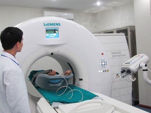
Chụp CT ổ bụng khi nào?
2. What does a CT scan of the abdomen show?
Abdominal CT scan results allow doctors to review and diagnose the following causes and conditions:Identify the cause of abdominal pain; Diagnosis of cancerous diseases of the abdominal organs, such as colon cancer, hepatocellular carcinoma and ovarian cancer; Check the status and extent of infection as well as abdominal trauma; Check the condition of the organs in some specific cases, including: gallbladder, pancreas, liver, biliary tract; Detect some kidney-related problems such as: kidney stones, pyelonephritis, hydronephrosis, ...
3. When is an abdominal CT scan indicated?
The patient is assigned to have an abdominal CT scan when the following symptoms appear:Abdominal pain and suspicion of abnormalities in the intestines; Bleeding during urination or defecation; Difficulty urinating, jaundice and unusual weight loss; Fever of unknown cause; Abdominal trauma; Detect abdominal fluid.
4. Abdominal CT scan procedure
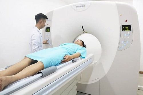
Just like the general CT scan procedure, patients assigned to have an abdominal CT scan will follow the steps below.4.1. Before taking the patient remove metal objects and jewelry from the body; The patient declares whether she is pregnant or not and some diseases related to veins, kidneys, asthma, diabetes and drug allergies; CT scan patients sign a commitment to inject contrast in case of mandatory use; Patients need to fast before the injection of contrast for about 4-6 hours. About 2 hours before the scan, the patient can still drink water as usual. Patients should take prophylactic drugs if there is a history of allergy before using contrast agent.

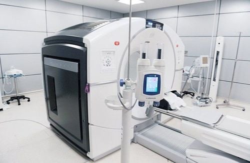
Tiêm thuốc cản quang trước khi chụp CT ổ bụng
5. Does CT scan of abdomen affect health?
Abdominal CT scan is a modern method to help diagnose the disease, but this method still affects health if used too much.Using X-rays with a dose of radiation passing through the abdomen is warned to be extremely dangerous for pregnant women and young children. Although the amount of X-ray used in CT scanning is committed to the acceptable level, if used regularly, the consequences are unpredictable. It may even have the potential to cause some cancers.
Therefore, medical experts recommend that, if not absolutely necessary, pregnant women and children should not prioritize this method.
If the patient is prescribed a CT scan, the doctor can ask the doctor about the amount of radiation used in the scan and the possible health risks before deciding to have the scan.
In general, abdominal CT is a relatively safe and low-risk imaging test, because the radiation exposure in each scan is adjusted so that the radiation dose is minimal. meet the image quality for doctors. In addition, with the development of increasingly modern science and technology, today, new imaging machines and equipment have been greatly improved to minimize the influence of X-rays on people. disease but still ensure diagnostic value.
Therefore, to be safe, in case the patient is a woman who is pregnant or suspected of being pregnant, it is necessary to inform the doctor to have a better alternative diagnostic method that does not affect the fetus.
CT scan is a method that brings many benefits and is prescribed by doctors to diagnose dangerous diseases. However, in order to have good and accurate diagnosis results, it requires doctors to have professional qualifications and experience, and to have modern equipment and machines.
Vinmec International General Hospital system across the country is invested with advanced technical facilities and a team of highly qualified and experienced doctors, which is a prestigious address for many patients to choose to visit. examination. The CT scan technique here will be performed according to standard procedures, giving the most accurate results, especially with the necessary considerations to ensure absolute safety for the patient.
Doctor Nguyen Le Thao Tram has many years of experience in the field of specialized imaging in pathology on ultrasound, radiology, multi-slice CT, magnetic resonance such as nervous system, spine, musculoskeletal system joints, soft tissue, tumor diseases, intra-abdominal inflammation and vascular diseases... In addition, Dr. Tram is also capable of performing fine needle aspiration (FNA) mammography, thyroid gland and subcutaneous soft tissue. Currently working at the Diagnostic Imaging Department of Vinmec Nha Trang International General Hospital.
Please dial HOTLINE for more information or register for an appointment HERE. Download MyVinmec app to make appointments faster and to manage your bookings easily.