This is an automatically translated article.
The article is professionally consulted by Master, Resident, Specialist I Trinh Le Hong Minh - Doctor of Radiology - Department of Diagnostic Imaging - Vinmec Central Park International General Hospital.Transesophageal echocardiography is still a new definition for many people, and makes many patients confused and concerned when they are assigned to perform this technique. So what is transesophageal echocardiography, indicated in what cases and contraindicated?
1. What is transesophageal echocardiography?
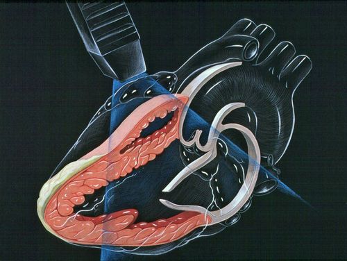
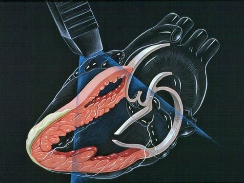
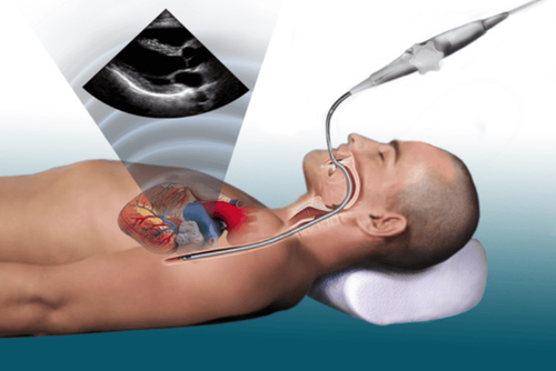
A transesophageal echocardiogram is a type of test that reconstructs your heart.In transesophageal echocardiography, sound waves play a very important role, they are responsible for recreating the most detailed images of your heart along with the blood vessels that originate or lead to the heart. Unlike conventional forms of echocardiography, the transesophageal echocardiogram transducer generates ultrasound waves and is attached by a very small tube, which enters through the mouth, then passes through the mouth. down the throat and into the patient's esophagus.
Because the esophagus is located very close to the heart chambers, we can completely obtain the most realistic and clear images of the structure and valves of the heart. Ultrasound waves in the form of esophageal ultrasound do not cause harm to the patient's health status, and can obtain much more detailed images than conventional ultrasound methods. from the outside.
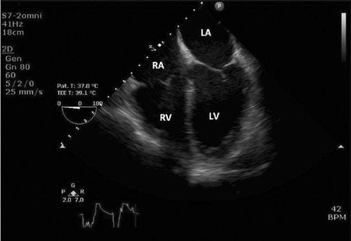
The main purpose of transesophageal ultrasound tests is to use ultrasound waves to create the most detailed images of the heart chambers, heart muscle, heart valves, along with the outer layer of the heart (also known as called the pericardium), and the blood vessels that are connected to the person's heart.
Usually, this method is usually indicated when the doctor wants to know more details about the structure of the patient's heart, after performing other routine ultrasound methods.
2. When is transesophageal echocardiography indicated?
Transesophageal echocardiography is a new ultrasound method that has been popular for several years now and offers much better results than older ultrasound methods, the resulting images are not only more specific but also much more detailed. .Doctors will ask the patient to perform a transesophageal echocardiogram in the following cases:
2.1 The patient has a transthoracic echocardiogram but the image is not clear The following are the main causes to this situation:
The patient is obese, or has recently undergone epigastric surgery, or is suffering from chronic lung disease. To look for atrial appendages, atrial thrombus (usually done before open heart surgery, mitral stenosis, or electric shock). Patients with infectious endocarditis (natural or prosthetic valves). Patients with congenital heart disease; Posterior structures (eg, pulmonary veins, main trunk or pulmonary branches, atria); Atrial septal defect; Coarctation of the aorta.
3. Contraindications to transesophageal echocardiography
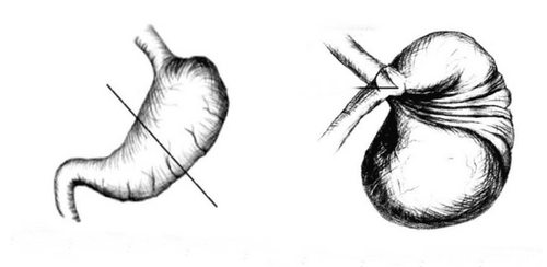
Absolutely contraindicated in the following cases:Lack of consent of the patient; The current patient is unwilling or uncooperative; Lack of technician expertise during TEE intubation; Esophageal obstruction (eg, patient with cancer, esophageal atrophy); Stomach torsion;
Accurate identification of esophageal disease; Varicose veins of the esophagus ; There is a diverticulum of the esophagus; Esophageal fistula; Patients with advanced esophagitis; Patients with carcinoma, scleroderma, gastric hernia; Open or closed thoracic esophageal trauma; History of esophageal surgery; Gastric reconstruction surgery to prevent gastric reflux in the esophagus; Abnormalities in the cervical spine: severe cervical osteoarthritis/patients with cervical spondylosis, or cervical spondylosis; Patients who have had neck surgery or radiation therapy to the neck; Severe deformities in the oropharynx; Radiotherapy for the first time in the mediastinum; Having internal bleeding. Before performing transesophageal echocardiography, the patient should fast for at least 5 hours. This will help the patient not have food reflux up the esophagus and at the same time do not have to experience discomfort and nausea during the ultrasound. In addition, patients should also abstain from alcoholic beverages or stimulants before the ultrasound.
Transesophageal ultrasound technology contributes to improving the effectiveness of treatment of complex cardiovascular diseases. Moreover, transesophageal echocardiography does not cause pain and discomfort to the patient, so you can rest assured to perform this ultrasound therapy at reputable hospitals across the country.
Before taking a job at Vinmec Central Park International General Hospital, the position of Doctor of Imaging Department since February 2018, Doctor Trinh Le Hong Minh used to work as a resident in the Radiology Department at hospitals: Cho Ray, University of Medicine and Pharmacy, Oncology, People's Gia Dinh, Trung Vuong... from 2012-2015. Officially worked at Cho Ray Hospital from 2015-2016, City International Hospital from 2016-2018.
Please dial HOTLINE for more information or register for an appointment HERE. Download MyVinmec app to make appointments faster and to manage your bookings easily.


