This is an automatically translated article.
The article was written by BSCKI Nguyen Viet Anh and MSc Tong Diu Huong - Department of Diagnostic Imaging, Vinmec Nha Trang International General HospitalThrough MRI images of the lumbar spine, it is possible to see whether or not the spinal canal narrows in the anterior and posterior direction, whether the spine degenerates to form bone spurs of the vertebral body...
1. About the lumbar spine
The lumbar spine consists of 5 vertebrae numbered from L1 to L5. Lumbar vertebrae are usually large, much stronger than the other vertebrae in the spine to bear the full weight of the human weight on it. The feature of this part is the cylindrical vertebral body, the small triangular vertebral foramen, the anterior/posterior dimension is larger than the horizontal. The dorsal segment of the spine is curved and concave posteriorly. Compared to the posterior, the anterior height of the vertebral body was greater in L1, L2, the medial segment at L3 and smaller in L4, L5.
2. What is lumbar spine magnetic resonance?
Magnetic resonance imaging (also known as MRI) is a technique that uses magnetic fields and radio waves to create detailed images of body parts. MRI is one of the imaging techniques doctors use to diagnose diseases in the lumbar spine in particular and other body parts in general.
The difference of MRI with X-ray, CT-Scan (computed tomography) is that it does not use radiation, so when taking an MRI, the patient will not need to worry about any biological effects. study or radiation. This is a non-invasive technique with no side effects. When taking MRI, it will obtain good anatomical detail, high contrast, can see most of the lumbar structures such as soft tissue, bone marrow signals of the vertebral body, components of nerves and nerve roots. meridians of the lumbar spine, joints, ligaments that other techniques such as X-ray, CT-Scan are limited.
But MRI will have limitations such as loud noise during the scan, a long scan time (30-60 minutes depending on the imaging technique), so for patients with claustrophobia or small children It can be very difficult to shoot as this technique requires lying motionless for a relatively long period of time.

Vị trí cột sống thắt lưng
3. How is this method done?
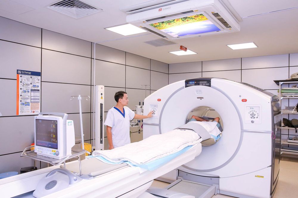
MRI helps detect a variety of diseases of the lumbar spine such as: herniated disc, vertebral spine, lumbar spine degeneration,... Not only that, it also identifies injuries such as spinal tuberculosis , there is a compression of a tumor, trauma, infection,... When you notice the following symptoms, you should think about consulting a doctor about an MRI scan of the lumbar spine: After carrying objects severe back pain or back pain for a long time with no known cause. Loss of bladder or bowel control. Have a history of known cancer, now have pain in the spine that is not relieved by pain medication. Numb limbs. Frequent pain spreading from the back to the buttocks, thighs and legs, affecting daily life. A typical MRI scan takes about 30-45 minutes. Patients do not need to fast, can change into simple clothes, do not bring anything on their body and arrange to lie neatly on the scanning table of the machine. Meanwhile, the patient can use headphones, both to reduce noise, both as a means of communication with outside technicians; while helping the patient to relax mentally. Only when relaxing the muscles of the whole body, the spine joints are kept in a fixed position, the image appears clearly. Keep the body in a fixed position because moving it will easily distort the results of the scan or make the image not clear, affecting the doctor's reading of the results later. The machine takes many shots, accompanied by noise, but the patient has been wearing ear protection so he will not feel any discomfort. Sometimes the person can feel a little heat but it won't be a big deal. If the patient has claustrophobia or is a young child, they will be advised to use sedation or anesthesia. The patient will be monitored for vital signs such as heart rate, breathing rate, blood oxygen level during the scan by doctors and anesthesiologists. Sometimes during the imaging process, it is necessary to use magnetic contrast medicine, the customer and/or the patient's family will be directly explained by the doctor about the diagnostic technical requirements, some risks affecting the disease status. or possible complications or allergic reactions, sign an undertaking before conducting the examination with contrast injection. Magnetic contrast allows the lesion to be prominent and helps the doctor better orient the lesion. The contrast medium used in MRI is generally safe, but as with all drugs, allergic reactions can occur.

Kỹ thuật chụp MRI cột sống thắt lưng được thực hiện tại cơ sở y tế uy tín
MORE:
Does Magnetic Resonance Imaging (MRI) have any effect? The role of magnetic resonance imaging (MRI) in the diagnosis of spinal and lumbar diseases Magnetic resonance imaging for diagnosis of spinal hernia


