This is an automatically translated article.
The article is expertly consulted by Master, Doctor Nguyen Thi Mai Anh - Doctor of Radiology - Department of Diagnostic Imaging and Nuclear Medicine - Vinmec Times City International General Hospital.Mammography is a method of screening for breast cancer in women. These scans may reveal breast abnormalities not found during physical examination.
1. Purpose of mammography
Mammography for cancer screening Mammography is a special breast imaging method that uses low-dose X-rays to record detailed images of the mammary glands for early detection of breast cancer. , even when there are no clinical symptoms, thereby helping patients to be treated at an early stage.Mammography to diagnose disease The doctor may order a mammogram to find the cause, explain the symptoms and identify the disease. Diagnostic mammography may be performed in the following cases:
Abnormal mass is palpable during physical examination Other symptoms are present: nipple discharge, newly appearing nipple retraction, breast tenderness, axillary lymph nodes, male gynecomastia... Biopsy, needle placement Diagnostic mammograms usually capture more breast images than screening mammograms.
2. Who will perform the mammogram for the patient?
The mammogram is performed by the technician and the radiographer will read the results.3. What do patients need to prepare before mammography?
Patients need to talk to their doctor or healthcare provider - talk about any symptoms you're having. If you are pregnant or breastfeeding, your doctor may delay your mammogram. The right time to have a mammogram is one week after your period ends. This is the time when the breasts are least sensitive. Notes to inform the breast implant doctor Have ever had breast surgery You are pregnant or breastfeeding Please exchange enough information so that the technician can take an accurate mammogram and the doctor Diagnostic imaging reads results more clearly.4. How is mammography done?
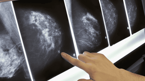
Chụp nhũ ảnh
The patient will place the breast on a flat surface. A sheet of plastic will be used to cover the top. The compression reduces the breast thickness so the radiation dose is reduced, holds the breast tightly to reduce image blur, and thins the mammary gland to separate the structures for better observation. Usually, for screening mammograms, the technician will usually take pictures of the breast in two positions: Craniolateral oplique (CC - Craniocaudal)) and medial lateral lopsided (MLO - Mediolateral oplique). When taking diagnostic mammograms, the patient will take two positions CC and MLO, if there is suspicion of injury, they can be taken with some other positions to get more information.
Pressure from breast compression may feel uncomfortable, but the sensation is fleeting and will help the radiologist to detect minor abnormalities and better read the results.
At Vinmec International General Hospital, all image data of mammograms as well as other mammograms are stored long-term on the PACS system. This system, as part of an electronic medical record, is convenient for monitoring and comparing lesions at subsequent visits to help improve diagnostic value.
Master, Doctor Nguyen Thi Mai Anh has nearly 10 years of experience in the field of diagnostic imaging, especially in imaging breast and thyroid cancer. Received formal training at Thai Binh Medical University and specialized postgraduate training at Hanoi Medical University. Currently, the doctor is a radiologist at Vinmec Times City International General Hospital.
Please dial HOTLINE for more information or register for an appointment HERE. Download MyVinmec app to make appointments faster and to manage your bookings easily.


