This is an automatically translated article.
The article is professionally consulted by Master, Doctor Le Hong Chien - Radiologist - Department of Diagnostic Imaging and Nuclear Medicine - Vinmec Times City International Hospital
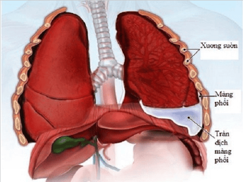
Pneumothorax or pleural effusion are common diseases of the respiratory tract. For treatment, the patient needs to have a pleural effusion or thoracentesis. Pleural sinus drainage is understood as a measure to release the compressed pleural space. However, to carry out this measure, it is necessary to have a doctor's appointment for pleural sinus drainage and follow specific procedures.
1. Place a pleural drain
Pneumothorax is a procedure to place a drainage tube into the pleural cavity. The purpose of this procedure is to drain harmful gases and fluids out of the body. The cause of pneumothorax is a punctured lung, which causes pain and difficulty breathing for the patient. Pleural effusion has many causes, patients will experience symptoms such as shortness of breath, lung pain when coughing. The procedure of inserting a pleural tube is a solution indicated by the doctor to intervene and free the pleural cavity from the compression of fluid or air.
Sinus pleural drainage is an invasive procedure, placing a drain through the chest wall. This procedure requires the operator to have expertise, technique and experience. At the same time, it is also necessary to ensure the principle of drainage during the implementation to bring optimal efficiency.

Thực hiện đặt ống dẫn lưu màng phổi khi bị tràn khí hoặc tràn dịch màng phổi
2. Pathological indications for pleural drainage tube placement
For some of the following cases, it will be indicated to drain the pleural cavity:
The patient has effusion, pneumothorax leading to the lungs being compressed, affecting respiratory function, causing difficulty breathing. Pleural effusion, pneumothorax recurred many times. The patient's pleura was filled with pus. After the injury, the patient's lungs appear to be overflowing with blood, or have complications after surgery. Effusion, hemothorax due to malignancies. The case of patients is indicated for artificial ventilation when the pleura is pneumothorax caused by trauma. Some cases of chronic pneumothorax, open pneumothorax, valve... Patients after applying the method of air aspiration, catheter placement failed.
3. Procedure for placing drains
In order to place a pleural sinus drainage tube, the doctors will proceed in turn according to the following procedure:
First, determine that the patient has completed all the necessary laboratory results such as: X-ray- chest x-ray, computed tomography scan, total peripheral blood cell analysis, blood coagulation function test and blood biochemistry...

Người bệnh thực hiện các xét nghiệm cận lâm sàng cần thiết trước khi tiến hành đặt ống
Check the patient's hemodynamic, respiratory and cardiovascular status, determine the ability to coordinate when placing a pleural tube. Instruct the patient into a semi-recumbent half-sitting position, with the head elevated at an angle of 30° and then administer pre-anesthetic injections if necessary. Determine the location of the pleural drainage tube: for pneumothorax, it is in the 4-5 intercostal anterior axillary line, for pleural effusion, it is in the 4-5 or 5-6 middle axillary intercostal space. Inject local anesthetic at the pleural opening area, administer anesthesia layer by layer, from the skin to the parietal leaf of the pleura, not letting the anesthetic come into contact with the vascular lumen. Use a specialized anesthetic needle to probe the pleural space. Using a surgical knife, make an incision in the upper border of the 6th rib about 0.5-1cm. Dissection of subcutaneous tissue with a pince without nodules, pointing the pince toward the superior border of the fifth rib. Using the hand to press on the intercostal muscles and the pleural wall, a sound can be heard when the tip of the pince touches the pleural space ( In case of pneumothorax). Open a hole just enough for the drain by separating the 2 pince ends. In the case of pneumothorax, bring the tube forward and above the midclavicular line. If it is a pleural effusion, the tube must be moved posteriorly and downward. The drain should be placed about 2-3cm deep in preterm infants. For full-term babies, after checking the status of gas or fluid discharge, the tube is placed about 3-4cm deep.

Trẻ sinh non tháng sẽ được đặt ống dẫn lưu vào sâu khoảng 2-3cm
Any questions that need to be answered by a specialist doctor as well as customers wishing to be examined and treated at Vinmec International General Hospital, please register for an online examination on the Website for the best service.
Please dial HOTLINE for more information or register for an appointment HERE. Download MyVinmec app to make appointments faster and to manage your bookings easily.