This is an automatically translated article.
The article is professionally consulted by Master, Doctor Nguyen Van Huong - Department of Diagnostic Imaging - Vinmec International Hospital Da Nang. The doctor has many years of experience in the field of diagnostic imaging.Pleural ultrasound has long been used to diagnose lung diseases from the chest wall, pleura, mediastinum to lung parenchyma. In particular, the strength of this imaging method is effusion diagnosis, pleural biopsy. So in what cases is the patient indicated for pleural ultrasound?
1. Advantages of pleural ultrasound
Pulmonary and pleural ultrasonography is an imaging method similar to other ultrasound techniques. Accordingly, the probe position is placed in certain positions to diagnose syndromes such as fluid in the lung parenchyma, pulmonary coagulation; pneumothorax and other pleural abnormalities.Therefore, pleural ultrasound is of great significance for the diagnosis and treatment of diseases related to the pleura and chest wall. Currently, lung ultrasound techniques are commonly performed techniques in medical facilities and hospitals.
Accordingly, pleural ultrasound has many outstanding advantages over other types of lung tests, specifically:
Cost: low cost of performing pleural ultrasound, suitable for all subjects. . High safety: The pleural ultrasound does not use any radiation like other imaging techniques, so it does not affect the patient's health. This is also the reason why multiple pleural ultrasounds can be performed. The process of performing pleural ultrasound quickly, with clear images and information at the survey ultrasound area, pleural ultrasound is considered to be more convenient and safer for patients than chest X-ray. In addition, the advantage of pleural ultrasound is that it does not cause pain, leaves no complications and the patient does not need to prepare anything before ultrasound like other methods.

Trắc nghiệm: Làm thế nào để có một lá phổi khỏe mạnh?
Để nhận biết phổi của bạn có thật sự khỏe mạnh hay không và làm cách nào để có một lá phổi khỏe mạnh, bạn có thể thực hiện bài trắc nghiệm sau đây.2. Disadvantages of pleural ultrasound
Besides the advantages, pleural ultrasound also has certain disadvantages, specifically:Resolution of the image depends on the device The image is not objective, depends on the person performing the ultrasound. Difficulty in analyzing some images, for example in the diagnosis of lung tumors. Since the lungs are the body's air reservoir, sound conduction is inhibited. The ultrasonic waves will be scattered by the air.

3. In which cases is indicated pleural ultrasound?
When suffering from respiratory diseases, depending on the severity, the patient has different symptoms and disease progression. The most common symptom of lung diseases is a cough. However, in some cases, coughing is a protective mechanism of the lungs that is not a disease cough. The cough reflex is the body's defense mechanism of the lungs when foreign objects enter. Coughing will push the foreign object along with the mucus out of the lungs quickly. But if the case of cough persists, the patient needs to perform imaging methods to identify lung diseases, at this time, pleural ultrasound can be prescribed by doctors. Specifically:Detect and evaluate pleural effusion Find out the cause of opacities in the chest wall or pleura (tumor, fluid) Differential diagnosis of pleural effusion or localized fluid under the diaphragm, evaluate Diaphragm mobility Detect and evaluate pleural thickening, pleural tumor, lung tumor invading the chest wall Guidelines for puncture, drainage, pleural biopsy Detect pneumothorax. In addition, when the patient has symptoms such as wheezing, shortness of breath, continuous cough, pain, discomfort in the chest area, appearance of sputum, the doctor may appoint the patient to perform a pleural ultrasound. to diagnose specific diseases.
4. How is pleural ultrasound done?
The pleural ultrasound is performed in 2 positions, specifically:Sitting position: Placing the transducer in the intercostal spaces, the doctor can examine the pathology of the chest wall, pleura, and find digital pleural effusion. little amount. Lying position: Examine the pleura at the lateral and posterior costal angles of the diaphragm, the lung parenchyma at the bottom, take the liver as an ultrasound window to evaluate the diaphragm and pleura.

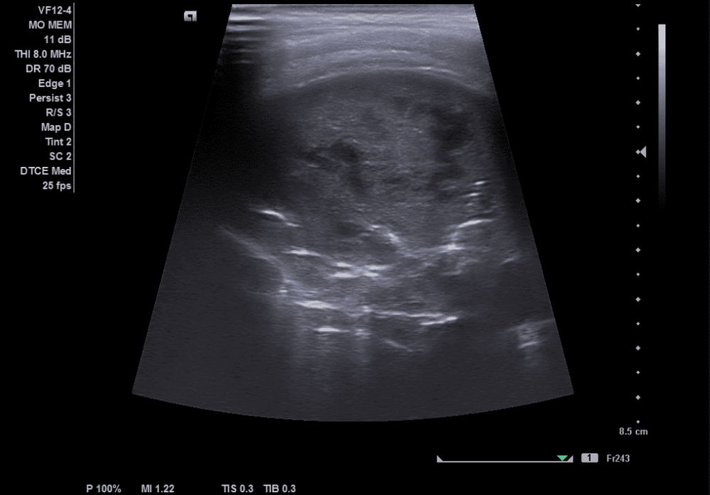
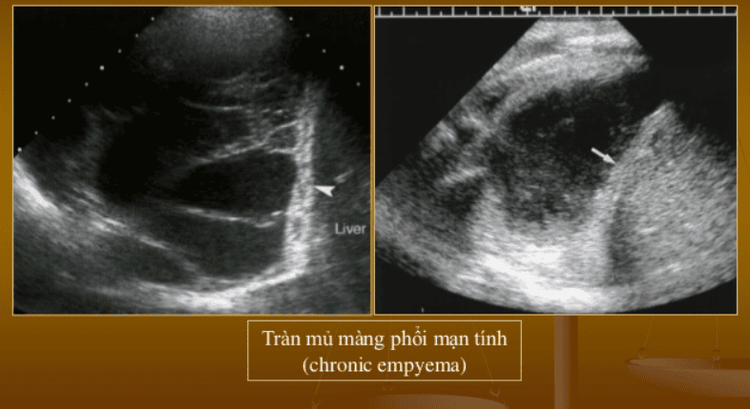
Step 1: Move the transducer along the intercostal position. If there is a lesion, compare with the contralateral side and observe the change of respiratory rhythms. Step 2: Observe the ultrasound image when the transducer moves to the position between the parietal pleura and visceral pleura, in case there is a negative space, this is a typical sign of pleural effusion. This step uses a 3.5 MHz probe to estimate the extent of the pleural effusion. Step 3: In the case of pneumothorax, the ultrasound image will not show the pleural slide, the comet tail and the enlarged pleural line. Pleural ultrasound has the ability to diagnose chest wall diseases such as trauma, pneumonia and many other respiratory diseases. When there is pleural fluid on ultrasound, depending on the amount of fluid and accompanying clinical symptoms of the patient, the doctor will require hospitalization or outpatient treatment. However, the results of pleural ultrasound depend greatly on the equipment and the sonographer. Modern equipment will give sharper, more detailed and specific images. From there, it is easy to detect abnormal signs even at an early stage with very small manifestations.
Currently, Vinmec International General Hospital has been using modern generations of ultrasound machines. One of them is GE Healthcarecar's Logic E9 ultrasound machine with full features, HD resolution transducers for clear images, accurate assessment of lung damage as well as many other organs. In addition, a team of experienced radiologists, trained in pleural ultrasound, in addition to assessing the amount of fluid and the nature of the fluid, can also detect small cases of fluid. and do pleural puncture procedures to drain the therapeutic fluid. Thanks to that, the diagnostic results are always accurate and bring high value, good support for the treatment process.
Please dial HOTLINE for more information or register for an appointment HERE. Download MyVinmec app to make appointments faster and to manage your bookings easily.


