This is an automatically translated article.
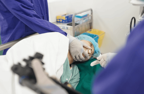
The article was professionally consulted by Specialist Doctor I Nguyen Thi Mai - Radiologist - Diagnostic Imaging Department - Vinmec Phu Quoc International General Hospital. Dr. Mai has many years of experience working in the field of diagnostic imaging.Gastrointestinal tract diseases are very common in clinical practice. Screening ultrasound is an accessible, noninvasive, non-radiative and inexpensive imaging modality, often chosen as the first-line diagnostic modality in gastroenterology, which avoids the routine use of surgical procedures. diagnosis is invasive and costly and leads to early implementation of appropriate treatment.
This is a promising method with high sensitivity and specificity, which has gained popularity in recent years and has the potential to become the method of choice in the diagnosis of many gastrointestinal tract pathologies.
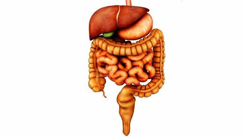
1. Digestive tube
The human alimentary canal is a linear system, interrelated in both structure and function. They come from the mouth down the pharynx to the esophagus, under each person's esophagus is the stomach and intestines (the intestines include the small intestine and the large intestine, while they are divided into different segments. The small intestine is divided into three segments: duodenum, jejunum, and ileum, while the large intestine is divided into three parts (cecum, colon and anus and anal canal) and the end of a normal digestive tract usually the rectum and anus.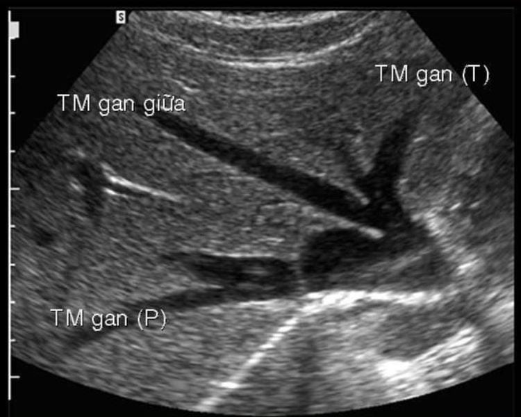
2. Ultrasound of the digestive tract
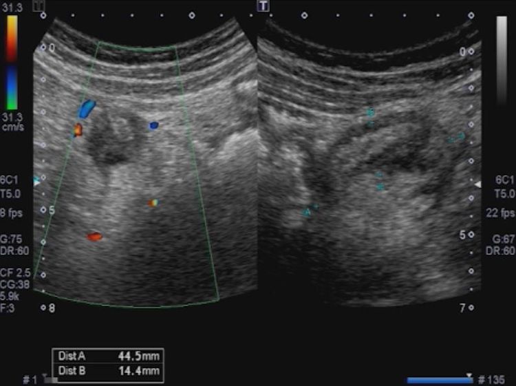
Some pathological abnormalities can be detected when performing ultrasound of the gastrointestinal tract and the ultrasound imaging characteristics of these cases are as follows:2.1 pyloric stenosis Children from 2 to 12 weeks of age, appear vomiting now. Longitudinal resection: pyloric thickness ≥ 3 mm, pyloric thickness ≥ 12 mm. Cross section: Target sign. Stomach: distention of fluid and/or gas. 2.2 Intussusception Intussusception is common between the ages of 3 months and 3 years. The most common type of ileocolic intussusception: the last segment of the ileum (intussusceptum) instils the colon (intussuscipiens). Formation of a bunion in the right abdomen, averaging 2.6 cm in diameter. The jejuno-jejunal intussusception has a mean diameter of 1.5 cm, in the left abdomen. 90% are idiopathic - idiopathic (due to mesenteric lymphadenitis), 10% have pathologies that create lead points - such as double bowel cysts, polyps... The frequency of recurrent intussusception is 10% after intussusception. Usually relapses within 48-72 hours. Ultrasound diagnosis of intussusception: + Cross section: Target sign.
+ Vertical cut: Sandwich sign (sandwish sign).
+ Can be seen in the bunion with mesenteric lymph nodes.
+Transient intussusception, usually jejunal intussusception.
2.3 Gastrointestinal tract inflammation Gastrointestinal tract inflammation is very common. Stomach, small intestine, large intestine, rectum: edema, increased perfusion in the gastrointestinal tract wall. Important: the wall of the digestive tract is symmetrically thick, without loss of layered structure. Fatty infiltrates (thick echoes) can be seen around the inflamed gastrointestinal tract. There may be free fluid in the abdomen. 2.4 Acute appendicitis Tubular structure, one-terminal, non-peristaltic, non-compressive, cross-sectional with target shape (outer diameter ≥ 7 mm, appendiceal wall ≥ 3 mm). McBurney sign on ultrasound (+). Fatty infiltration around the appendix (thick echogenic). Appendiceal stones: found in obstructive appendicitis. When stones are present, the risk of perforation increases. Less fluid around the appendix. The tip of the appendix is often the site of initiation of infection and perforation. 2.5 Colonic Diverticulitis Normally colonic diverticula are barely visible on ultrasound. The diverticulum can be in the sigmoid colon, the left colon, or sometimes the right colon.
Diverticulitis :
Always associated with Colitis. Air in the diverticulum creates a dirty back shadow. Fat infiltration around an inflamed diverticulum. Increased perfusion of the infected area. - In addition, it is also possible to detect lesions such as: Inflammation of the colonic fat mane, tumors of the digestive tract.
3. Conclusion
Gastrointestinal ultrasound is increasingly being developed and widely applied in the examination and treatment of gastrointestinal tract diseases. can detect abnormal images, so that diseases can be detected early, especially serious diseases, and have timely treatment.Please dial HOTLINE for more information or register for an appointment HERE. Download MyVinmec app to make appointments faster and to manage your bookings easily.


