This is an automatically translated article.
The article was professionally consulted by Specialist Doctor II Nguyen Quoc Viet - Department of Medical Examination & Internal Medicine - Vinmec Danang International General Hospital. The doctor has more than 20 years of experience in the examination and treatment of cardiovascular diseases and Cardiovascular Interventions (Including angiography, dilation, stenting of coronary arteries, renal arteries...), placing temporary pacemakers , forever...Hypertrophic cardiomyopathy is a disease in which the left ventricular wall and interventricular septum become abnormally thick, making it harder for the heart to pump blood. The disease is usually diagnosed by the patient's examination and echocardiography for other reasons, or when sudden death due to arrhythmia occurs. However, in a small number of people with left ventricular hypertrophy, the thickened heart muscle can cause shortness of breath and chest pain on exertion, but has not been properly examined and treated.
1. What is left ventricular hypertrophy?
Hypertrophic cardiomyopathy is an abnormal thickening of the left ventricular wall that cannot be explained by other causes of cardiac overload. The disease is usually detected based on echocardiography, computed tomography (CT scan) or magnetic resonance imaging (MRI) of the heart when measuring the thickness of the left ventricular septum from more than 15 mm.In contrast to other cardiovascular diseases, left ventricular hypertrophy often occurs in young people, in athletes and is one of the causes of sudden cardiac death when it has not been detected before.
The classification of left ventricular hypertrophy plays a decisive role in the prognosis of the disease progression as well as the treatment orientation for the patient. Accordingly, left ventricular hypertrophic cardiomyopathy is classified in many ways as follows:
According to hemodynamics: Left ventricular hypertrophic cardiomyopathy causes obstruction when increasing the maximal gradient across the left ventricular ejection chamber at rest from above 50 mmHg before blood enters the general circulation. In contrast, left ventricular hypertrophy is not obstructive if the pressure at this site is less than 30 mmHg.
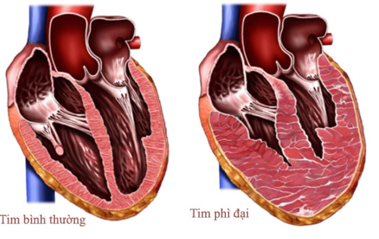
Left ventricular anterior septal hypertrophy: accounting for 10% Hypertrophy of the anterior septum and posterior wall: accounting for 20% Hypertrophy of the entire anterior and posterior ventricular septum and lateral wall: accounting for 50% Other areas of the left ventricle are larger than the anterior and posterior walls of the septum: 18%.
2. What are the symptoms of left ventricular hypertrophy?
The first manifestation of left ventricular hypertrophy is diastolic heart failure when the patient finds it difficult to breathe on exertion, at night when lying down or has angina even at rest. If left ventricular hypertrophic cardiomyopathy causes obstruction, even patients often experience dizziness or fainting, especially during exertion.Examination of patients with left ventricular hypertrophy shows strong apex, palpable carotid pulse and 2-peak pulse waves while the gradient is normal. Auscultation with galloping sounds with T4 common and T3 uncommon. In obstructive hypertrophic cardiomyopathy, an ejection systolic murmur is heard in the aortic socket and if the Valsalva maneuver or squatting is performed, the murmur will increase in intensity.

3. How to test and diagnose left ventricular hypertrophy?
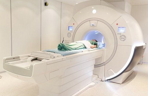
The diagnosis of left ventricular hypertrophy is usually based on cardiac imaging. In which, the most commonly used tool is echocardiography with the following characteristics:Signs of left ventricular hypertrophy: left ventricular mass index over 110g/m2 in women or over 134g/m2 in men Thickness of the ventricular walls: normally the thickness of the heart walls is less than 11mm, in typical cases, the upper wall is 15mm; Usually asymmetric LV thickening with a posterior wall/wall thickness ratio of 1.3 to 1.5. Ejection chamber obstruction: based on maximum pressure gradient across the ejection chamber from more than 30 mmHg SAM sign: anterior mitral leaflet movement during systole Decreased left ventricular diastolic diameter Attention in cases of ventricular wall If the left is less dense, such as with only 13 or 14 mm on ultrasound, a combination of clinical and other imaging modalities is required before ruling out the diagnosis. In addition to echocardiography, cardiac MRI is indicated when the ultrasound image is not clear, especially the lateral basal and left ventricular apex or the SAM sign. On the other hand, cardiac MRI is also of great significance in clinical practice because the maximum wall thickness if greater than 30mm will be a major prognostic factor of the disease associated with sudden arrhythmic death. Simultaneously, MRI is also needed to differentiate from other causes of left ventricular hypertrophy such as Fabry disease and amyloidosis.
In addition to diagnostic tests, genetic testing for hypertrophic cardiomyopathy should be considered in individuals with a clear phenotype of hypertrophic cardiomyopathy to facilitate screening and management. to other family members. Currently, more than 20 genes that cause hypertrophic cardiomyopathy have been detected, however, if the family members have negative gene tests, they still have to periodically monitor the heart every 6 to 12 months through symptoms. electrocardiogram, echocardiography and echocardiography.
4. How to treat left ventricular hypertrophic cardiomyopathy?
The general principle of the treatment of left ventricular hypertrophy should be achieved is to improve the patient's functional symptoms by eliminating the outflow obstruction and improving the elasticity of the left ventricle. Besides, treatment of arrhythmias, prevention of dangerous arrhythmias can cause sudden death. Not only that, the treatment of left ventricular hypertrophy is also aimed at preventing and treating major complications such as infective endocarditis, thromboembolism, and sudden death.Left ventricular hypertrophy is also treated with common cardiovascular drugs because no specific drug has been found so far. Drugs that can be used prophylactically are beta-blockers or calcium channel blockers to reduce disease progression as well as contribute to symptom improvement, reducing the increase in left ventricular ejection fraction gradient that occurs with exercise. In addition to drugs, the treatment of left ventricular ejection fraction obstruction can also be carried out with measures such as surgery to remove the ventricular septum or the procedure to thin the interventricular septum with alcohol through the heart catheter.
For the prevention of the risk of sudden arrhythmic death, the patient will have an indication for defibrillator placement when one of the following risk factors is present:
Ever received cardiopulmonary resuscitation Recurrent syncope Occurrence of tachycardia ventricular electrocardiogram with continuous monitoring or electrophysiological exploration Significant left ventricular thickening with interventricular septum greater than 30 mm Blood pressure does not increase with exercise Family history of sudden death. In addition, as soon as a patient with left ventricular hypertrophy is detected, it is necessary to conduct an early echocardiogram for all their parents and siblings. If these subjects are children and adolescents, ultrasound should be performed every 3 years, then every 5 years when reaching adulthood.
In summary, left ventricular hypertrophy is a rare cardiomyopathy and cause of sudden death that is not detected before conduction disturbances occur. Accordingly, the above information will help contribute to early disease detection, active treatment as well as screening plans for other family members, to prevent unfortunate events that may occur.

Please dial HOTLINE for more information or register for an appointment HERE. Download MyVinmec app to make appointments faster and to manage your bookings easily.


