This is an automatically translated article.
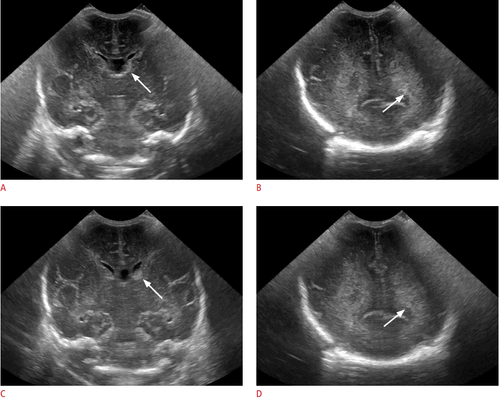
The article is professionally consulted by MSc, BS. Dang Manh Cuong - Doctor of Radiology - Department of Diagnostic Imaging - Vinmec Central Park International General Hospital. The doctor has over 18 years of experience in the field of ultrasound - diagnostic imaging.Ultrasound is a method that uses high-frequency sound waves to create an image of a part of the inside of the body. Ultrasound scans can be used to monitor an unborn baby, diagnose a medical condition, or direct surgeons in certain pathologies.
1. Types of ultrasound
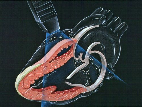
Ultrasound is a method of using high-frequency sound waves through a transducer to create images of the inside of the body or the organs and organs that the person appointing the ultrasound needs to observe. Ultrasound is often used to monitor the growth of the fetus, detect abnormalities in the abdominal organs, pelvis, muscles, heart and blood vessels.... Ultrasound is usually performed externally. body (non-invasive ultrasound) however in some cases the ultrasound probe will be inserted into some natural opening of the body such as the vagina to observe the female genitalia or the esophagus to check. Check the heart and some blood vessels or also rectally in case a rectal exam and prostate exam are needed.Depending on the purpose of the doctor and the organ to be examined, people divide ultrasound into 3 main types: Non-invasive ultrasound, invasive ultrasound and endoscopic ultrasound.

Invasive ultrasound: In many cases doctors An internal ultrasound will be required to get a clearer picture of certain organs such as the prostate, ovaries or uterus... This is also a form of transrectal ultrasound. The typical form of ultrasound of this type is the transvaginal and anal ultrasound. The patient is asked to lie on their back or side, depending on the case, with their knees towards the chest. A small, sterile probe (condom or latex) is gradually inserted by the technician into the vagina or anus and an image is displayed on a screen. An internal ultrasound may cause some discomfort to the patient, but this should clear up by the end of the ultrasound. This form of ultrasound is usually done quickly and takes little time.

Usually, before the ultrasound, the patient will be given an anesthetic and numb the throat because endoscopic ultrasound can bring a very uncomfortable feeling. In addition, the patient also protects the mouth and teeth, avoiding the case of biting the endoscope.
2. Types of ultrasonic waves
2.1. Definition of Ultrasound An ultrasonic wave is a sound wave with a frequency greater than 20kHz. They travel at the speed of sound. Therefore, their wave length is less than 333200cms-1/20000Hz = 1.66 cm(ג = v / υ).
2.2. Types of Ultrasonic Waves Ultrasonic waves can propagate through a medium as medium, strong, or intense waves, depending on the elastic properties of the medium. Based on the particle displacement of the medium, ultrasonic waves are classified into the following four types:

3. Types of ultrasound machines
On the market today, there are many types of ultrasound machines, diverse in types, uses, forms, quality and different prices. Here are some common types of ultrasound machines:
Please dial HOTLINE for more information or register for an appointment HERE. Download MyVinmec app to make appointments faster and to manage your bookings easily.


