This is an automatically translated article.
The article is professionally consulted by MSc, BS. Dang Manh Cuong - Doctor of Radiology - Department of Diagnostic Imaging - Vinmec Central Park International General Hospital. The doctor has over 18 years of experience in the field of ultrasound - diagnostic imaging.Swollen lymph nodes in the neck are a common, sometimes normal sign of an inflammatory response in the body. But it can also be a serious medical condition like melanoma. Cervical lymph node ultrasound is the simplest method for early detection of abnormal lymph nodes.
1. What is cervical lymph node ultrasound?
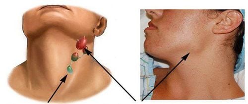
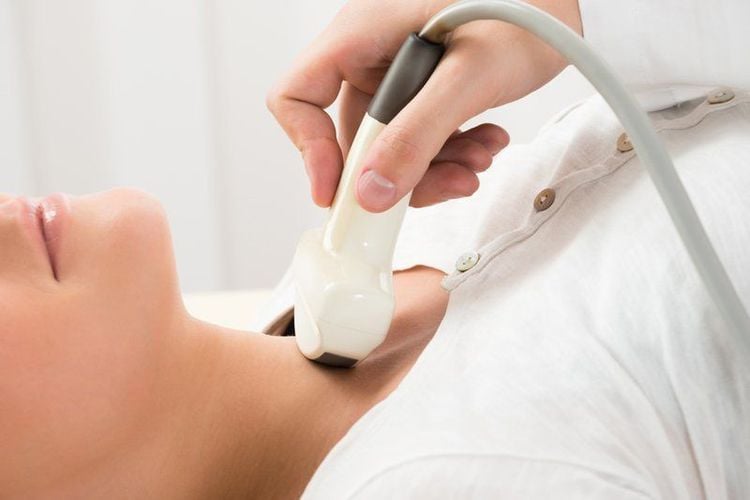
Neck lymph nodes are the lymph nodes in the neck that function in the immune response against pathogens. Lymph nodes are smooth, oval or flattened structures located along the path of the lymph node.Cervical lymph node ultrasound is the use of high-frequency ultrasound waves to image the internal structures of the cervical lymph nodes. Through examination, the position, size, shape, and internal structure of the cervical lymph nodes can be observed. From there, it can help detect abnormal signs of cervical lymph nodes, thereby making a diagnosis or orienting the diagnosis of the disease if in doubt.
The cervical lymph node ultrasound method is a simple, non-invasive method that does not cause discomfort to the patient, but has high diagnostic value.
2. Indication method of cervical lymph node ultrasound
Cervical lymph node ultrasound is indicated when there are any abnormal signs such as:Patient can feel the lymph nodes in the neck such as submandibular lymph nodes, angled nodes, supraclavicular nodes... Self-observation has many floating nodes. in the neck. When touching the lymph nodes in the neck, they feel a lot of pain, feel a lot of pain at night. The skin of the neck lymph nodes is red, bruised, sore... Swollen lymph nodes accompanied by signs such as fever, fatigue, weight loss, loss of appetite... Combined with swelling of other lymph nodes in the body. Ultrasound to guide the localization of aspiration cytology for tissue biopsy or cytology. Cervical lymphadenopathy may be a sign of a normal inflammatory response but may also be a sign of a malignancy that should be ruled out. Therefore, early lymph node examination is very meaningful in diagnosing, providing appropriate treatment, and ensuring patient's health.

3. How is the cervical lymph node ultrasound performed?
3.1 Preparations Performers: Radiologists, technicians. Means: Ultrasound machine with flat transducer, high frequency above 7MHz, ultrasound machine should be used in combination with color doppler to examine more effectively; specialized gel used in ultrasound; cloth to wipe the patient after the ultrasound. Patients: No special preparation is required because eating and drinking does not affect the ultrasound results, the diagnostic ultrasound method is clearly explained for the patient to cooperate with. Patients need to provide full information related to the disease to the doctor. 3.2 Procedures Patient position: Lying on the back, with both hands raised above the head or under the nape of the neck, the patient needs to tilt his or her neck back to expose the neck area to be examined by ultrasound.Step 1: Apply a specialized gel to the neck lymph nodes. Using an ultrasound probe to scan groups of lymph nodes in the neck region, it is necessary to continuously scan the patient's skin to avoid missing lesions. Step 2: When examining the lymph nodes, it is necessary to describe the location of the lymph node group and the number of abnormal nodes. The shape of the lymph node, the border of the affected lymph node needs to be examined. Evaluation of lymph node structure (compared with neighboring muscle structures): Hyperechoic, hypoechoic or homophonic, see if there are calcified nodules inside the lymph node, lymph nodes have signs of fluidization, necrosis... Measure lymph node size : It is necessary to measure the horizontal diameter, the longitudinal diameter, the ratio between the horizontal and vertical diameters to help orient the risk of malignancy, the thickness of the lymph node shell... Determine the organization of the hilar node: Is the hilar node missing and the thickness not? even the nucleus pulposus. Doppler ultrasound of the lymph nodes: To evaluate the vascularity of the lymph node cortex or the center of the lymph node. It is necessary to survey the adjacent organs in the cervical lymph node when detecting cervical lymphadenopathy: It is necessary to further investigate the patient's thyroid and salivary glands when abnormal lymph nodes are present in the neck, especially when there are lymph nodes. supraclavicular... Step 3: Evaluation of results Normal: The lymph nodes are small in size, short in diameter, appearing at locations such as under the jaw, at the angle of the jaw, in the posterior triangle of the neck; The hilar nodes are often clearly seen on ultrasound. Malignant lymphadenopathy: Large lymph nodes are seen, appearing in abnormal positions in the neck (superclavian lymph nodes), often round in shape, irregularly thickened, may not clearly see the hilar nodes, often hypoechoic, there may be calcifications in the lymph nodes, peripheral vascular proliferation...Malignant tumors with lymph node metastasis or lymphomas causing changes in lymph node structure. Inflammatory lymph nodes: Usually large in size, small in number, oval or flattened, hilar lymph nodes can be seen, and central lymph node vasculature may be enlarged... This is also a benign condition, encountered during an inflammatory response to a viral infection. common creatures. Step 4: Review the ultrasound results and print the results. Indicates additional laboratory methods to confirm the diagnosis if necessary. Cervical lymph node ultrasound can detect a number of diseases based on the location, size, number and shape of cervical lymph nodes. However, the accurate assessment of the pathological condition depends on the experience of the radiologist. Therefore, for the best and most accurate results, it is necessary to visit reputable medical facilities to limit false diagnoses and affect the treatment process.
Please dial HOTLINE for more information or register for an appointment HERE. Download MyVinmec app to make appointments faster and to manage your bookings easily.


