This is an automatically translated article.
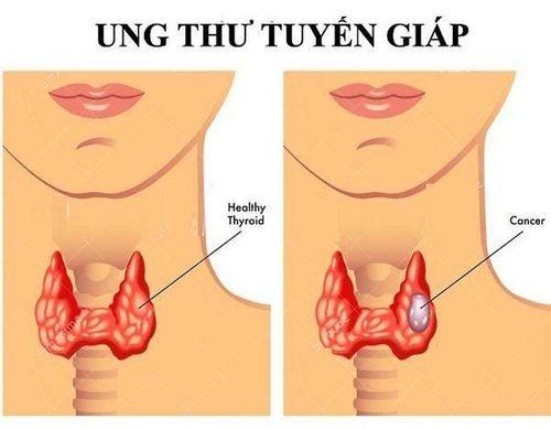
The article is professionally consulted by Master, Doctor Le Hong Chien - Doctor of Radiology - Intervention - Department of Diagnostic Imaging and Nuclear Medicine - Vinmec Times City International General Hospital.The thyroid gland is an endocrine gland located in front of the trachea consisting of 2 lobes connected by a thyroid isthmus. Ultrasound is a simple method that plays an important role in the assessment of thyroid pathology, especially diffuse thyroid disease.
1. What is diffuse thyroid disease?
Diffuse thyroid disease refers to thyroid conditions that cause enlargement of the gland, which can affect thyroid function.
Thyroid disease includes a number of diseases such as: simple goiter, thyroiditis, thyroid cancer, basedow's disease...
Diffuse thyroid pathology is very diverse, the diagnosis of the disease is not based on the medical conditions. Clinical manifestations, subclinical signs such as ultrasound, laboratory tests, biopsies... are essential for definite diagnosis of the disease.
In which, ultrasound is a simple, cheap, easy to perform and non-invasive method to help support the diagnosis. In addition, in malignant thyroid disease, the combination with ultrasound elastography is very meaningful in diagnosis and limits unnecessary aspiration or biopsy for patients.
2. Ultrasound in diffuse thyroid disease
The role of ultrasound in diffuse thyroid disease is to determine the nature and nature of the lesion mass. From there, orient or diagnose thyroid disease.2.1 Thyroid goiter pathology There can be a single or multinodular goiter. When examining the ultrasound, it is necessary to determine the gland volume, distribution of goiter, shape, size, structure and influence of the goiter on the muscles. nearby organs.Ultrasound images often show irregular enlargement of the thyroid gland, with many nodules appearing. Assess whether the nodule is hyperechoic or hypoechoic. The majority of uniform, hyperechoic hypertrophy are usually benign. On the contrary, if the negative density in the nuclei is irregular, hypoechoic, microcalcified nodules appear, and angiogenesis is increased, it may be a malignancy.
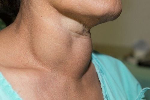
Hình ảnh bệnh nhân mắc bệnh lý bướu giáp
2.2 Basedow disease Thyroid ultrasound is a simple method to help orient and diagnose basedow disease.
When basedow pathology ultrasound shows images like:
The thyroid gland is larger than normal, the thyroid isthmus is thicker. The thyroid gland has a diffuse, heterogeneous hypoechoic structure, and small hypoechoic foci can be seen. When using vascular doppler, thyroid angiogenesis was found. Basedow's disease is often accompanied by signs such as shaking hands, increased body temperature, rapid pulse, bulging eyes... So it can be combined with clinical signs to accurately diagnose the disease.
2.3 Thyroiditis Some inflammatory thyroid diseases can be encountered such as De Quervain's thyroiditis, autoimmune chronic thyroiditis (Hashimoto's disease), Riedel's thyroiditis, and acute purulent thyroiditis.
Hashimoto's thyroiditis: + Acute stage: Usually, the ultrasound image is diffuse hypoechoic throughout the thyroid gland and cause gland enlargement, accompanied by vascular proliferation.
+ Chronic stage: The disease usually develops for many years, the ultrasound image is diffuse hypoechoic image, the gland size can be normal or atrophy, without vascular proliferation.
De Quervain's thyroiditis: The disease is less common than Hashimoto's thyroiditis, presenting clear clinical signs. Ultrasound shows bilateral asymmetrical lesions, often localized, located at the edge of each lobe. Riedel's thyroiditis: The ultrasound image shows a diffuse band of fibrosis in 1 or both lobes and spreads into surrounding tissues. Acute purulent thyroiditis: A rare condition, on ultrasound, an abscess is seen with the manifestation of a solid fluid, clearly demarcated with healthy tissue, with a crust that can see air inside the abscess. .
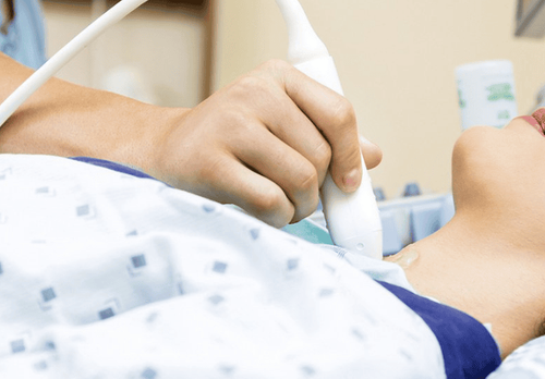
Siêu âm giúp bác sĩ chẩn đoán xác định một số bệnh lý tuyến giáp
2.4 Thyroid cancer Thyroid cancer is a rare type of cancer, more common in women than men and in all age groups. Increased in subjects who often irradiate the neck area.
When doing ultrasound, it is necessary to evaluate the organization of the lesion, especially the thyroid tissue around the gland and the status of cervical lymph node metastasis.
Ultrasound images of thyroid cancer often show hypoechoic or mixed-negative masses, rarely homophonic masses, uneven lesion margins, and angiogenesis.
Papillary cancer: The most common type and also the least dangerous. Ultrasound shows hypoechoic mass, when elastography shows hard tissue, there may be calcifications or microcalcifications in the mass. May be in the form of warts in the wall of the cyst, can see cervical lymph node metastasis. Adenocarcinoma: Ultrasound image is hypoechoic mass, cystic condition is rare, lymph node infiltrates are rare, and there is no microcalcification. In general, ultrasound findings are not specific, so diagnosis should be based on aspiration cytology or tissue biopsy. Undifferentiated cancer: Common in the elderly, the prognosis is very poor. On the ultrasound image, the thyroid gland has a large mass, surrounding infiltrates and infiltrates on lymph nodes. Lymphoma and secondary cancer: Secondary cancer of the thyroid gland is uncommon. The ultrasound image is a hypoechoic, or mixed, multi-nodular image.
Currently, thanks to the development of science and technology, people apply elastic ultrasound of thyroid tissue to orient the diagnosis of thyroid cancer with higher accuracy. Help patients limit unnecessary aspiration or biopsy.
Lesions in the thyroid gland that cannot be seen with the naked eye can be detected through ultrasound. In addition, thyroid ultrasound is also said to be the simplest method to evaluate and monitor pathology in the thyroid gland.
Therefore, as soon as you feel abnormalities in the thyroid gland, the patient should examine and conduct an ultrasound of the thyroid gland to detect problems in the thyroid gland early. In addition, we should also proactively go to the doctor and periodically monitor to detect health abnormalities early for timely treatment.

Định kỳ khám và kiểm tra tuyến giáp giúp phát hiện sớm những vấn đề tại tuyến giáp
Currently, Vinmec International General Hospital has an effective screening and screening for thyroid diseases, helping to check thyroid function and early detection of common thyroid diseases such as: simple goiter pure, hyperthyroidism, hypothyroidism, thyroiditis, thyroid nodules, thyroid cancer, etc. to have the best treatment measures to ensure your own health.
The examination process at the hospital always meets international standards with a team of qualified doctors, a modern and civilized medical examination and treatment environment. Therefore, customers can rest assured.
Please dial HOTLINE for more information or register for an appointment HERE. Download MyVinmec app to make appointments faster and to manage your bookings easily.