This is an automatically translated article.
The article is expertly consulted by Master, Doctor Tong Van Hoan - Emergency Medicine Doctor - Emergency Department - Vinmec Danang International Hospital1. Hemothorax- pneumothorax in closed chest trauma
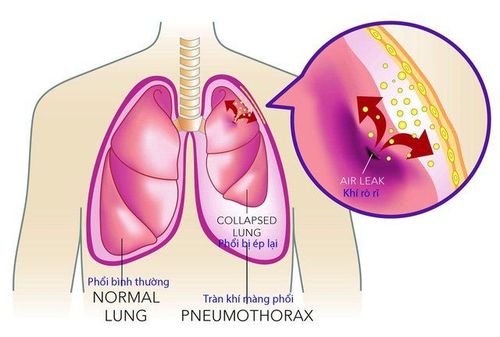
Closed chest trauma is an injury that damages the chest wall and organs in the chest, but the chest wall is still closed, the pleural cavity is not open to the outside air. Closed chest trauma is usually caused by the chest wall being struck by a blunt object or the chest being pressed by two hard objects. Closed chest trauma directly affects breathing and circulation, the patient can die in a short time, so it needs urgent emergency care.Hemothorax - pneumothorax is one of the most common forms of blunt chest trauma, occurring in over 85% of common thoracic injuries. Under normal conditions, the pleural cavity is an air-free space, with a negative pressure of -10 mmHg to -12 mmHg. Negative pressure helps the lungs expand during respiration. In closed chest trauma, air enters the pleural space usually from the tear of the lung parenchyma, blood enters the pleural space from the tear of the lung parenchyma, the rib fracture, and damage to the internal organs of the chest. Blood is in the low zone, qi is in the high zone. Blood and air will occupy space and press on the lung parenchyma, causing a loss of negative pressure in the pleural space, causing atelectasis, the mediastinum will be pushed to the opposite side.
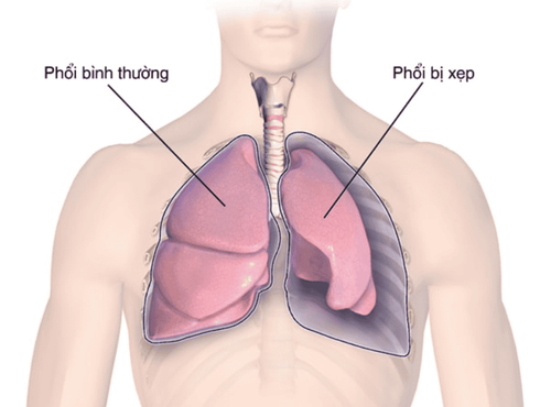
When a traumatic hemothorax occurs to a greater extent than a pneumothorax, the clinical and radiographic findings resemble a simple hemothorax. Caused by small punctured lung parenchyma, causing less air to enter the pleura, or ruptured large blood vessels of the chest wall, causing a lot of bleeding.
2. Diagnosis of hemothorax- pneumothorax
Diagnosis of hemothorax - pneumothorax is based on clinical symptoms and laboratory findings.Clinical symptoms:
Patient has difficulty breathing and chest pain continuously, increasing gradually. With severe trauma, shortness of breath and chest pain will appear immediately after the injury. With mild lesions, symptoms will appear late, when the amount of blood and air in the pleural space is large enough to cause symptoms. In mild form, patients rarely have systemic signs. But when hemothorax-pneumothorax is severe, the patient will have respiratory distress symptoms such as cyanosis of the lips and extremities, rapid pulse, reduced oxygen saturation SpO2, and sweating. There may be manifestations of anemia such as blue skin, pale mucous membranes, rapid pulse, cold limbs, sweating. Physical signs in the chest and respiratory apparatus include: Decreased respiratory amplitude on the injured side of the chest, the patient has rapid shallow breathing, has symptoms of rising and falling of the nose, pulling the respiratory muscles in the neck and chest. when inhaled.

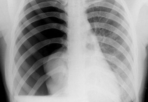
X-ray film shows clearly displaced ribs, subcutaneous emphysema, and the center is pushed to the healthy side. If the film is taken in the upright position, the typical hemothorax-pneumothorax is a low-level hemothorax, separated from the high-grade sign by a horizontal line. Blood tests: white blood cells are elevated, there may be signs of anemia. Computed tomography: shows clear images and hemothorax-pneumothorax, based on CT scan can assess the extent and have the right attitude to handle these patients. Pleural ultrasound: there is blood in the pleural space.
3. Treatment of hemothorax- traumatic pneumothorax
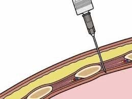
After receiving the patient, quickly clear the airway and give the patient oxygen. Actively resuscitate, administer fluids, and give blood transfusions in case of hemorrhagic shock. Give the patient pain reliever, antibiotic, tetanus shot if there is a scratch. Then, transfer the patient to the operating room. If the patient has severe pneumothorax, it is necessary to insert a needle into the pleural space and subcutaneous tissue to relieve pressure, then transfer the patient immediately to the operating room.
Minimal drainage of the pleural space: Anesthesia with local anesthesia, drainage through the intercostal space 5-mid axillary line. If the pneumothorax is large, an additional airway may be placed through the 2-way midclavicular intercostal space. When draining immediately blood > 1500ml within 6 hours after injury or follow-up after drainage see blood >200ml for 3 consecutive hours. Consider thoracotomy for hemostasis of large vascular lesions of the chest wall and lung parenchyma. If there is a lot of air drainage, the lungs do not expand, and the hemodynamics do not improve, the doctor may prescribe a thoracotomy to stitch the pleural-bronchial tears. After surgery, patients will be instructed to practice respiratory therapy to avoid atelectasis, use antibiotics, pain relievers, anti-inflammatory drugs, cough suppressants to prevent infection and reduce symptoms. Most patients with hemothorax-pneumothorax in blunt chest trauma have a good prognosis if treated aggressively and correctly.
Pneumothorax is a fairly common medical condition today with many causes. In order to have the right and reasonable treatment, the patient needs to be diagnosed accurately thanks to imaging techniques. modern. Currently, Vinmec International General Hospital is one of the leading prestigious hospitals in the country, trusted by a large number of patients for medical examination and treatment. Not only the physical system, modern equipment: 6 ultrasound rooms, 4 DR X-ray rooms (1 full-axis machine, 1 light machine, 1 general machine and 1 mammography machine) , 2 DR portable X-ray machines, 2 multi-row CT scanner rooms (1 128 rows and 1 16 arrays), 2 Magnetic resonance imaging rooms (1 3 Tesla and 1 1.5 Tesla), 1 room for 2 levels of interventional angiography and 1 room to measure bone mineral density.... Vinmec is also the place to gather a team of experienced doctors and nurses who will greatly assist in diagnosis and detection. early signs of abnormality in the patient's body. In particular, with a space designed according to 5-star hotel standards, Vinmec guarantees to bring patients the most comfort, friendliness and peace of mind during the treatment of pneumothorax at the Hospital.
Please dial HOTLINE for more information or register for an appointment HERE. Download MyVinmec app to make appointments faster and to manage your bookings easily.


