This is an automatically translated article.
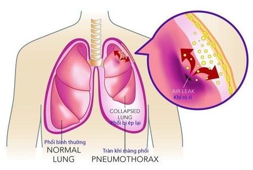
The article was written by Master, Doctor Tran Thi Diem Trang - Department of Examination & Internal Medicine - Vinmec Central Park International General HospitalPneumothorax is a condition in which air builds up between the lungs and the chest wall. Air entering the pleural space can come from the lungs or from outside the body. If not treated promptly, pneumothorax leaves dangerous complications.
1. Pneumothorax
Pneumothorax is divided into 2 types, divided according to its cause, including:
However, the tear usually occurs at the site of the parapleural air bubbles. Small air bubbles may appear near the pleura, the walls of which are not as strong as the normal lung parenchyma and may cause tearing. The air then exits the lungs but is trapped between the chest wall and the lungs.
Most cases of primary pneumothorax occur in healthy young adults without any pulmonary disease, being more common in tall and thin individuals.
Around 2 out of 10,000 young adults in the UK develop a primary spontaneous pneumothorax each year. Pneumothorax is more common in men than in women. In people over 40 years of age, spontaneous pneumothorax is rare. It is also more common in smokers than in non-smokers. Tobacco smoke seems to make the walls of the air bubbles weak and prone to tearing.
About 3 in 10 people with primary pneumothorax will have more than one recurrence after treatment at some point in the future. Pneumothorax occurs usually on the same side and usually occurs within the first three years.
1.2 Secondary spontaneous pneumothorax Secondary pneumothorax means a pneumothorax that is a complication of an existing lung disease. If lung disease weakens the pleura, pneumothorax will occur. A weakened pleura makes it easier for the pleura to tear and allow air to escape from the lungs.
Therefore, pneumothorax can be a complication of chronic obstructive pulmonary disease (COPD). Due to chronic obstructive pulmonary disease, air bubbles develop more. In addition, other lung diseases can also complicate pneumothorax, including: pneumonia, sarcoidosis, cystic fibrosis, pulmonary tuberculosis, lung cancer, and idiopathic pulmonary fibrosis.
1.3 Other causes of pneumothorax Other causes such as trauma to the chest can cause pneumothorax. Even chest surgery can cause pneumothorax. Pneumothorax is also a rare complication of endometriosis.
Chest X-ray can confirm pneumothorax. If a pneumothorax is suspected to be caused by a lung disease, your doctor may order additional tests.
At Vinmec International General Hospital, chest radiographs are conducted with fully digital X-ray machines (Digital Radiography). This reduces radiation dose by up to 50% compared to previous “traditional” X-ray machines. Thereby ensuring maximum safety and efficiency in the diagnosis and treatment of diseases.

2. Possible complications of pneumothorax
Recurrence: Nearly half of people with a history of pneumothorax experience a relapse, usually within the first three years. Air is constantly leaking: Even though a drain has been placed to draw the air out, sometimes air can continue to leak. After a few days to a week or so, you may need surgery to repair the leak. Lack of oxygen: Pneumothorax causes compression of the lungs, less air entering the lungs leads to less oxygen entering the blood. Lack of oxygen can lead to a lack of oxygen supply to the organs and in severe cases can be life-threatening. Cardiac tamponade: If not treated early, pneumothorax will cause cardiac tamponade. Pneumothorax can affect blood flow to the heart. Cardiac tamponade can be fatal if not treated immediately. Respiratory failure: occurs when the oxygen level in the blood drops too much. Severe hypoxia can lead to cardiac arrhythmias and disturbances of consciousness such as unconsciousness, confusion, somnolence, and coma. Ultimately, respiratory failure can be fatal. Shock: This is quite a serious condition, occurring when blood pressure drops very low and the vital organs of the body are deprived of oxygen and nutrients. Shock is a medical emergency and requires immediate care.3. Treatment of pneumothorax

3.2 It is sometimes necessary to drain the accumulated air If the pneumothorax is larger or if the patient has pre-existing lung disease or difficulty breathing, drain the accumulated air. In the event of a tension pneumothorax, prompt drainage is imperative.
The most common method is to insert a very thin drainage tube through the chest wall with the guidance of a needle. A large syringe with a three-way valve will be attached to the small tube to be threaded through the chest wall. After air enters the syringe, turn the three-way valve to expel the air in the syringe. This will be done until the air in the pleural space is removed.
In cases of secondary pneumothorax with associated lung disease, a larger tube is required through the chest wall to drain the large pneumothorax. Usually, a pneumothorax is placed for a few days to allow the torn lung tissue to heal.
3.3 Recurrence after treatment Some people have spontaneous pneumothorax that occurs repeatedly. About 3 in 10 people with primary pneumothorax will have more than one relapse after treatment at some point in the future.
The doctor will then advise on prevention, for example surgery is an option for the patient if a pleural tear and air leak have been identified. The doctor will inject an irritating powder, usually a talcum powder, onto the surface of the lungs. This will cause inflammation, but then help the surface of the lung to stick better to the chest wall.
People who have had a pneumothorax should avoid deep diving, because pressure changes can lead to a risk of not being able to breathe. Do not fly without pleural drainage. If you regularly dive or fly, for example those who work in the aviation industry, are pilots or cabin crew, or are professional divers, special measures may be required and may have to be taken. pleural adhesions. Smoking cessation is necessary to reduce the risk of recurrent pneumothorax.
Pneumothorax can leave dangerous complications, which can recur after treatment. Therefore, patients need to adhere to the principle of treatment, avoiding pressure changes. When you see any unusual signs such as chest pain, shortness of breath, etc., you need to go to a medical facility for timely examination and intervention.
Please dial HOTLINE for more information or register for an appointment HERE. Download MyVinmec app to make appointments faster and to manage your bookings easily.


