This is an automatically translated article.
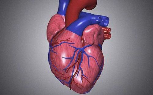
The heart is the first organ to form and function. This highlights the importance of transporting matter to and from the developing embryo into the fetus. The heart's formation begins around day 18 or 19 from the dermis and begins to beat and pump blood around day 21 or 22.
1.The meaning of the formation of the heart
The first system to develop due to the increasing metabolic demands of the embryo is the cardiovascular system. Initially by simple diffusion of nutrients, necessary and sufficient but eventually becomes insufficient to supply oxygen and nutrients to the constantly growing embryo.
Accordingly, the formation of the heart is a complex interplay, which ensures proper structural formation and spatial configuration change in the appropriate time. Any influence that interferes with this process, whether genetic or environmental, leads to the development of congenital heart disease.
Thus, understanding the formation of the embryonic heart is necessary to understand the pathophysiology of congenital heart disease. The scientific community's knowledge of cardiovascular embryology and congenital heart disease has greatly expanded. The understanding of genetic influences at different stages of development has given a deeper insight into the management of congenital heart disease. In general, the embryonic development of the heart is very tightly regulated but is still influenced by genetics as well as environmental influences.
2. What is the anatomical process of heart formation?
Heart progenitors and arterial progenitors Embryonic development of the cardiovascular system begins with the migration of cardiac progenitor cells in the epiphysis, right next to the primitive line. These progenitor cells eventually develop into cardiomyocytes. In this same endoderm of the dermis, the so-called "islets", eventually undergo a stage of angiogenesis to form vascular structures. The aggregation of blood islets eventually forms an area known as the cardiovascular field. The cardiac field is initially horseshoe-shaped and surrounded by cardioblasts with the apex of the cardiac field, which eventually develops into the primitive ventricle with their respective egress pathways. Finally, the cardiac field changes configuration, forming a primitive cardiac tube that is continuous with surrounding vascular structures.

Quá trình hình thành của quả tim theo giải phẫu
The head aspect of the cardiac duct directs blood into the dorsal aorta, while the caudal aspect serves as a conduit for systemic venous drainage back to the heart. The closing of the neural tube and the anterior displacement of the pharyngeal membrane occur simultaneously to facilitate the migration of the embryonic heart into the thorax. The primitive heart tube consists of three layers, similar to the adult heart. The endocardium forms the endothelial layer of the embryonic heart. The myocardium forms the muscular mass of the embryonic heart while the visceral pericardium forms the outer surface of the embryonic cardiac duct.
Division of preliminary structures in the heart On around day 22-23, the heart tube lengthens and changes configuration, forming a heart ring. The cardiac ring forms when the apical aspect of the cardiac duct curves horizontally and to the right while the caudal aspect of the cardiac duct curves dorsally and to the left. The ring formation process takes about five days and is usually completed by day 28.
The different sections of the heart tube develop in the following pattern:
The proximal portion of the cardiac duct develops into the grooved portions of the right ventricle .
The middle segment of the cardiac duct is the glial and is the precursor to the ventricular ejection chambers.
The distal portion of the heart duct is called the trunk duct, leading to the proximal portion of the aorta and pulmonary artery.
Before the end of the formation of the great arterial root, the cardiac duct is essentially smooth-walled. Near the end of the loop formation, however, grooved regions begin to form, serving as primordial ventricles.
The septum usually forms between day 27 and day 37 through the fusion of tissue masses. These masses of tissue are called endocardial cushions and contribute to the formation of the atrial septum, interventricular septum, channels, and valves. By the end of the 4th week of development, the roof of the common atrium develops a crest-like structure called the septum, which is crescent-shaped. The two lower extremities of the septum move toward the endocardial cushion. Because the septum and endocardial cushion do not fuse completely initially, an opening remains, known as the primary foramen.
The endocardial cushions eventually fuse with the septum. Physiological autolysis creates perforations in the septum, which eventually coalesce to form a structure called the secondary foramen. This opening allows blood from the right primordial atrium to the left primordial atrium. Eventually, right atrium enlargement occurs, in which a new cardiac septum develops in the right atrium but never completely divides the atrium. This cardiac septum forms an incompletely overlapping crescent configuration with the primary septum. The remaining opening is the foramen ovale, one of two fetal structures responsible for directing blood flow from the developing lung (the other being the ductus arteriosus).
Formation of cardiac chambers At the end of the 4th week of pregnancy, the atrioventricular canal develops two atrioventricular cushions along the superior and inferior contours and two lateral cushions. The superior and inferior endocardial cushions eventually project into the lumen and eventually fuse, although the lateral cushions do not participate in this process. The result is the division of the atrioventricular canal into two distinct holes (left atrioventricular canal and right atrioventricular canal). Mesenchymal tissue surrounds the peripheral edges of each atrioventricular canal. This mesenchymal tissue eventually thins to form the atrioventricular valves. The valves themselves connect to the thickened papillary muscles through ligaments. During the fifth week of development, nodules will appear on the inner surface of the heart. The top right note moves towards the left, while the bottom left note moves towards the right. These bulges eventually rotate around each other to merge, forming the aortic septum, which divides the heart into two channels: the aortic and pulmonary channels. Simultaneously, the outflow tracts of the right and left ventricles are also formed. The development of the semilunar valve also begins to complete.
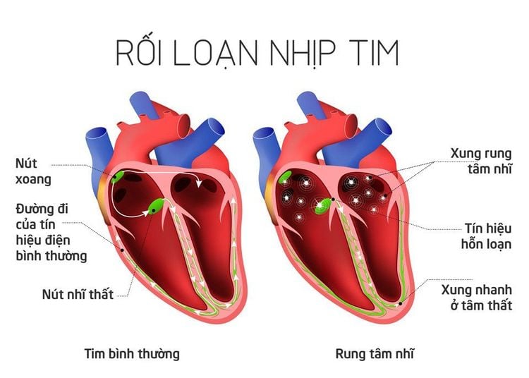
Các yếu tố Shox2 có thể dẫn đến rối loạn nhịp tim chậm bẩm sinh
Pacemaking The pacemaker of the primitive cardiac duct is located in the caudal end. Later in development, the sinus veins will take over the role of regulating the heart rhythm of the embryonic heart. Its incorporation into the right atrium serves as the origin of the sinoatrial node.
Studies have determined that the sinoatrial node, the atrioventricular node, and the proximal bundle branches have different origins. Although many genetic factors influence the structural development of the heart, several factors play a role in the development of the conduction system itself. In particular, Shox2 factors have been speculated to be able to lead to abnormal cardiac phenotypes, especially congenital bradycardia.
Embryonic Heart Function The cardiovascular system is the initial system to develop due to the increasing metabolic demands of the embryo. Compared with the cardiovascular system of adults, when the pulmonary system is well developed in adults, the embryonic heart function has many differences to help adapt and function well in the infancy.
In the amniotic fluid environment, the fetus will not need a developing lung system (because the placenta acts as a medium for oxygen exchange). Certain cardiovascular structures (such as the foramen ovale and ductus arteriosus) minimize blood flow to the fetal lungs by bringing blood into the circulatory system and out of the lungs. Minimal blood flow to the developing lung system prevents early lung development. The ductus arteriosus is a small blood vessel that connects the pulmonary artery and aorta, allowing blood flow from the right ventricle into the systemic circulation, bypassing the immature fetal lungs. Prostaglandins maintain the ductus arteriosus, and it closes soon after birth. The remnants of this structure are arteriolar ligaments. Premature closure of the ductus arteriosus is usually caused by pharmacological agents that block prostaglandin synthesis (ie, NSAIDs).
Birth shows an interesting change in fetal cardiovascular function. The fetus is no longer in the amniotic fluid environment and now needs the lungs to function to meet its respiratory needs - the first breath of oxygen a newborn takes in increases the induced oxygen tension, enabling blood flow to the lungs. Increased blood flow to the lungs leads to increased blood flow to the left atrium, which closes the foramen ovale.
In summary, the human heart is the first functional organ to develop, starting to beat and pump blood around day 21 or 22, just three weeks after fertilization. This highlights the importance of the heart in distributing blood through the blood vessels and exchanging nutrients, oxygen, and waste, important for both the developing fetus and out of its growing body. increasing very quickly. Accordingly, understanding the formation of the heart is the foundation for the study of the mechanism, diagnosis and treatment of congenital heart diseases later.
The formation of the fetal heart is a sign of the existence of the fetus in the womb, a special period in pregnancy. Accordingly, between 18 and 20 weeks, the mother can feel and hear the baby's heartbeat. At this time, the mother needs to pay attention to the fetal heart rate, regular and scheduled fetal ultrasound to check the cardiovascular status as well as the child's development, in order to detect birth defects early and have appropriate methods. early treatment.
Vinmec International General Hospital offers a comprehensive maternity care program for pregnant women right from the first months of pregnancy with full antenatal check-ups, periodical 3D and 4D ultrasounds and routine tests to ensure that the mother is healthy and the fetus is developing comprehensively.
Pregnant women will be consulted and checked for health under the close supervision of experienced and specialized Obstetricians, helping mothers to have more knowledge to protect their health during pregnancy as well as reduce pregnancy loss. reduce complications for mother and child.
Please dial HOTLINE for more information or register for an appointment HERE. Download MyVinmec app to make appointments faster and to manage your bookings easily.


