This is an automatically translated article.
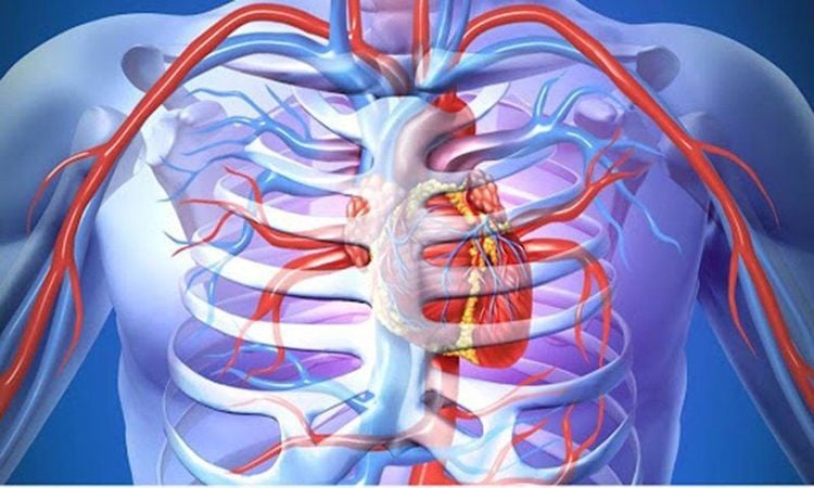
Open chest wounds are a group of common surgical emergencies. Depending on the anatomical lesions in the thoracic cavity, open chest wounds have many diseases with different names, severity - mild and different treatment and emergency.
1. Overview of open chest wound
The cause of an open chest wound is usually sharp objects (knife, scissors, iron rods) pierced into the chest. Sometimes, the wound opening is not on the chest wall but from the neck down or the abdomen up. Open chest wounds are common in men, aged 20 - 40.
1.1. Types of Open Chest Wounds In chest wounds, there are many different types of lesions, depending on the location, cause and characteristics of the wound. In fact, depending on the number and combination of types of lesions, they have different names. Some main types of injury include:
Injury to the chest wall
Perforation of the chest wall: A foreign body penetrates the chest wall, causing the pleural cavity to communicate with the outside. Through the stoma, air and blood flowing from the wound will enter the pleural cavity, causing symptoms of hemothorax - pneumothorax, signs of hemoptysis - gas, possibly subcutaneous emphysema, loss of pressure negative force,... If the wound is not sealed in time, the patient may die; Fracture, broken ribs: When penetrating the chest wall, foreign bodies can cause partial or complete rupture of the rib body in one or more bones, rupture the intercostal vessels, cause bleeding into the pleural space, otherwise Timely hemostasis can be fatal. In some cases, the wound penetrated the sternum or sternum rib, causing cardiac injury, rupture of the internal mammary vessel bundle, causing heavy bleeding into the pleural space or causing large mediastinal hematoma; Diaphragm perforation: May occur when the chest wound is located at the level of the intercostal space 5. Perforation of the right diaphragm is often accompanied by a right liver injury; Perforation of the left diaphragm is often accompanied by injuries to the left liver - spleen - stomach - colon - splenic angle. Patients often have bleeding - pleural fluid a lot, can have diaphragmatic hernia. Injury to the pleural cavity
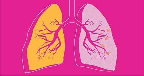
Hemothorax - pneumothorax: is a lesion that always exists as a constitutive element of an open chest wound. Air and blood enter the pleural space from the wound. Air in the pleural space can pass through the chest wound and cause subcutaneous emphysema around the wound; Pleural blood clots: When large amounts of blood flow into the pleural space at a rapid rate, part of the blood will clot into blood clots. At this point, only thoracotomy or laparoscopic surgery can be done to clear the blood in the pleura. If pleural drainage alone is not possible, these clots cannot be aspirated, causing pleural coagulopathy. Injury to organs
Alveolar or small bronchial laceration: Is a persistent lesion, a constitutive element of an open chest wound. Because the parietal and visceral pleura are very thin and close to each other, a foreign body that punctures the parietal leaf will perforate the visceral and lung parenchyma, cause bleeding, and release air into the pleural space; Tear of the great bronchi: A rare injury in an open chest wound, if it occurs, it is very serious because the wound penetrates deeply into the chest and mediastinum, causing complicated damage; Atelectasis: Common in blunt chest trauma but rare in chest trauma. manifestations of atelectasis are the collapsed alveoli, the lungs cannot expand, do not exchange gases and can cause many serious consequences; Heart and Pericardial Wounds: Heart injuries are common when the wound opening is located in a dangerous area of the heart. Depending on the size of the large or small pericardial perforation, there are different conditions. If the perforation is large, it often causes acute blood loss, leading to death after the injury. If the perforation is small, blood cannot escape from the pericardial cavity in time, and clots inside, which helps to temporarily seal the heart wound but causes acute cardiac tamponade. In addition, there may be an intermediate form of the above two conditions; Injuries to the aorta, thoracic aorta, and major blood vessels of the neck: Rare but very serious injuries caused by acute blood loss or into the pleural space causing massive hemothorax or out perforation in the chest wall. Anatomical injury in simple open chest wound
Chest wall perforation; Hemothorax - pneumothorax; Fracture - broken ribs; pleural coagulation; Tear of lung parenchyma; Lung collapse.

Nguyên nhân gây vết thương ngực hở thường là do các vật nhọn đâm vào ngực.
1.2 Diagnosis of open chest wound Physical signs: Shortness of breath and continuous, increasing chest pain; identify the cause of the injury (knife, scissors, slash or sharp object). Systemic signs: Mild: If the amount of blood - gas in the pleural cavity is not much, the chest wound is sealed, the general condition is little changed; Moderate or severe: If the chest wound is open or there is a lot of hemothorax, the patient has respiratory failure, blood loss or both. Respiratory failure includes rapid pulse, purple skin and mucous membranes. Symptoms of blood loss include rapid pulse, blue skin, pale mucous membranes, if severe blood loss may shock (drowsiness, sweating, cold hands and feet, low blood pressure,...). Thoracic sign: Wound: The entry hole is above the chest wall or below the ribs or base of the neck (or even distal to the ribcage). The wound can be in closed or open form, the size can be a few centimeters small or larger than 10 centimeters; Signs of hemothorax - pneumothorax syndrome: Chest deformity, nasal flaps, retraction of respiratory muscles in the chest and neck, rapid shallow breathing, murmurs, reduced or absent lung alveoli at the lesion site, puncture the pleural cavity on the injured side (indicated when there are clinical signs and the chest X-ray image is not clear) showing pneumothorax or hemothorax; Subclinical signs: Straight chest X-ray: Identify and evaluate the extent of injuries such as broken ribs - broken ribs, hemothorax - pneumothorax. If necessary, it is possible to take a lying position if the patient's condition does not allow it; Hematology tests showed signs of anemia, increased white blood cells; Pleural ultrasound showed fluid in the pleural space; Chest computed tomography is indicated in special cases (seeing hemothorax - pneumothorax, pleural blood clot, pulmonary - pleural foreign body).
2. First aid and treatment of open chest wounds
As the cause of serious disorders of respiratory and circulatory physiology, which can lead to death, open chest wounds are the first priority surgical emergency in first aid, diagnosis and treatment. In addition, because the open chest wound has many different types of injury, if it is not indicated correctly or treated in time, with good postoperative care, it can cause many complications and severe sequelae for the patient. pleural clots, empyema, pleural deposits, thickening of the pleura), affecting the health and working capacity as well as the economy of the patient.
2.1 First aid for an open chest wound Quickly seal an open chest wound: Usually using a compression bandage with a thick layer of gauze. If the wound is too large, stitches can be used to temporarily seal the wound. The Depage button is only performed when the wound is too large and there is no condition for suturing the wound; Clear airway, give oxygen if possible; Resuscitation, fluids and blood transfusion if the patient is in hemorrhagic shock; For patients to use antibiotics, pain relievers (the Paracetamol family, preferably intravenous form), tetanus vaccination; Quickly bring the patient to a surgical facility capable of operating on open chest wounds.

Vết thương ngực hở là loại cấp cứu ngoại khoa được ưu tiên hàng đầu trong sơ cứu, chẩn đoán và xử lý
2.2 Treatment of open chest wounds The treatment techniques for open chest wounds depend on the extent of the injury, but are mainly: Intervention to help the patient restore respiratory physiology (pleural drainage, suturing the wound) chest wall) and hemostatic intervention (if there is a cause for heavy bleeding).
Preparation
Implementation personnel: Surgeon; Basic facilities: Pince, scissors, drains of all sizes, sutures; Patients: Be clearly explained about the medical condition and risks of surgery, agree and sign a written commitment to surgery; Estimated time: 120 minutes. Carrying out surgery
Patient position: The patient lies on his back or on his side depending on the position of the wound, raising his hands behind his neck; Anesthesia: Local anesthesia, regional anesthesia or anesthesia depending on the extent of the wound; Technique: Suture wound: Antiseptic skin; clean the wound with water, hydrogen peroxide and Betadine; filter out corrupted software organizations, if any; suture the wound with Vicryl Dafilon 3.0 thread; Pleural drainage: Locate the V intercostal space in the middle axillary line or the second intercostal fissure in the midclavicular line -> disinfect the skin -> spread the skin -> 3cm skin incision -> wait sutures, fix the thread even though it is saved - > Use Pince to separate through the chest wall muscle layers into the pleural cavity -> place a 32F Silicon drain to drain blood, 28F to drain air -> rotate the drain in all directions to get all blood and fluid -> fix the guide save -> install a continuous suction system of -20cm H2O. Follow-up after surgery
Monitor the amount of blood and gas out according to the drainage; take care of drainage, ensuring the requirements of sterility, tightness, unidirectionality and continuous suction; monitor complications; Treatment of complications: Accidents related to drainage location: Drainage is low close to the diaphragm, drainage is under the skin, drainage is too deep, drainage into the abdomen. Handling depending on specific situations; Bleeding: If there is bleeding at the site, apply pressure and stitches to strengthen the skin edge; if hemothorax, drainage a lot, need to open chest to handle; Injury to abdominal organs: Need to open the abdomen to handle; Purulent pleurisy: Need thoracoscopy or thoracotomy for management. Emergency and treatment of open chest wounds need to be done quickly and according to instructions to help patients recover soon and minimize the risk of complications and unwanted sequelae.
Please dial HOTLINE for more information or register for an appointment HERE. Download MyVinmec app to make appointments faster and to manage your bookings easily.


