This is an automatically translated article.
Posted by Master, Doctor Nguyen Le Thao Tram - Doctor of Diagnostic Imaging - Department of Diagnostic Imaging - Vinmec Nha Trang International General Hospital
Accessory breasts, polymastia, supernumerary breasts, or mammae erraticae is a condition in which a person has at least one extra breast. The accessory mammary gland may be present with the nipple or areola. This is a relatively common congenital condition in which abnormal extra breast tissue is present alongside normal breast tissue.
1. Causes of extra mammary glands
Polycystic breast condition often occurs during fetal development. Normally, breast tissue will grow along the milk line and the extra tissue will disintegrate and be absorbed into the body. Multiple breasts occur when additional tissues do not disintegrate before birth. This condition can be inherited.

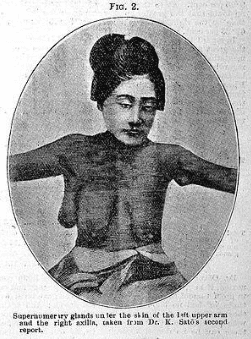
Hình 1. Phụ nữ với nhiều tuyến vú phụ
2. Location of the accessory mammary gland
This normal variant can present as a mass anywhere along the path of embryonic breast tissue (axillary to inguinal region). In which, common sites include armpits (60-70%), chest wall, vulva, knees, side of thighs, buttocks, face, ears, neck.
3. Expression of extra mammary glands
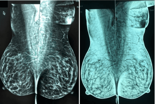
This condition occurs in 2-6% of women and 1-3% of men. Although the extra mammary gland is present from infancy, most people aren't aware of it until puberty. Most women only know they have extra breasts when they are discovered incidentally on an ultrasound or mammogram.
Common manifestations of accessory mammary glands include discomfort, pain, lactation, armpit thickening, and local skin irritation. Extra breast tissue responds to hormonal stimulation and may become more apparent during menstruation, pregnancy or lactation. Size varies from a small cluster to a complete structure (may include the nipple and areola).

Hình 2: Tuyến vú phụ trên hình chụp nhũ ảnh
4. Pathology of the accessory mammary gland
Literature in the world has described benign and malignant diseases of the accessory mammary glands in adult women. In which, benign pathology includes fibroids, mastitis or changes in fibrous sheath. Secondary mammary glands have a higher propensity to develop malignancy and occur at an earlier age.
However, the good news is that breast cancer in the accessory mammary gland only has an incidence rate of about 6%. The most common pathology is invasive ductal carcinoma, with approximately 50%-75%. Excessive breast growth (macromastia) can be seen during pregnancy as well as during adolescence.
5. Image characteristics and survey means
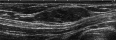
Most mammograms are found incidentally on routine mammograms. The ultrasound image shows breast tissue, indistinguishable from normal breast tissue. Occasionally, mammography is performed in atypical cases. Manifestations of signal and enhancement properties are similar to those of normal glandular tissue.

Hình 3. Hình ảnh siêu âm nách phải cho thấy khối 2,5 cm, hình bầu dục, khối song song với da có vách ngăn, tăng sinh mạch máu nhẹ. Sinh thiết lõi dưới hướng dẫn siêu âm cho kết quả bệnh lý u tuyến vú.
![Hình 4. Hình cộng hưởng từ tuyến vú phụ vùng nách trái (vị trí khung ”]” ).](/static/uploads/20200617_014034_484853_screenshot_15923578_max_1800x1800_png_d913868307.png)
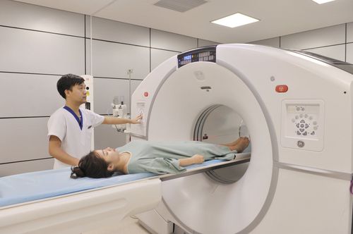
Hình 4. Hình cộng hưởng từ tuyến vú phụ vùng nách trái (vị trí khung ”]” ).
6. Treatment
Most cases of extra mammary glands do not require treatment. However, when symptoms are present, the treatment of choice is surgical excision, which reduces physical discomfort or mechanical discomfort in cases of large mammary glands.
To register for examination and treatment at Vinmec International General Hospital, you can contact Vinmec Health System nationwide, or register online HERE
REFERENCES
Ersilia M. DeFilippis1 and Elizabeth Kagan Arleo, “The ABCs of Accessory Breast Tissue: Basic Information Every Radiologist Should Know”, American Journal of Roentgenology. 2014;202: 1157-1162. 10.2214/AJR.13.10930. Bickle I. and Radswiki, Assessory breast tissue, https://radiopaedia.org/articles/accessory-breast-tissue. Ruqayya Naheed Khan et al, “Invasive carcinoma in accessory axillary breast tissue: a case report”, Int J Surg Case Rep, 2019, 59: 152-155.


