This is an automatically translated article.
The article is professionally consulted by Dr., Doctor Tran Nhu Tu - Head of Diagnostic Imaging Department - Vinmec Danang International General Hospital.
Although radiation causes only tissue damage and increases the risk of cancer when exposed to large doses, the radiation risks from imaging tests such as X-rays or CT scans should still be considered. be taken care of.
1. Radiation risks in daily life
First, it is necessary to properly understand the nature of radioactivity as artificial radiation, so these two terms are considered to be the same definition of a similar substance. Even the average healthy person is constantly exposed to radiation from a number of sources on a daily basis, including radioactive substances in the environment, indoor radon, and radiation from outer space. This is called background radiation and varies by geographic location. In which, 1 microsievert (mSv) is a unit of measurement of radiation exposure.
According to a study in the United States, the average American is exposed to about 3 mSv radiation from natural sources each year. However, background radiation exposure levels vary from state to state across the country as well as around the world.
Radon, the name for a natural gas found in homes, is the largest source of background radiation, the human body receives about 2 mSv per year. Radon concentrations are not the same across locations within the same country.
Because the earth's atmosphere can block some cosmic rays, geography also plays a significant role in the background radiation map. This means that people living in higher places increase their exposure to radiation. The radiation exposure of people living in high altitude states like New Mexico and Colorado (USA) is about 1.5 mSv/year more than people living near sea level as an example. A 10-hour flight also increases passengers' exposure to cosmic rays by about 0.03 mSv.

Tìm hiểu và trao đổi với bác sĩ trước khi thực hiện phóng xạ
2. Radiation sources from diagnostic imaging exams
The amount of radiation received from imaging tests depends on the type of examination, as well as on which part of the human body needs to be diagnosed. Specifically:
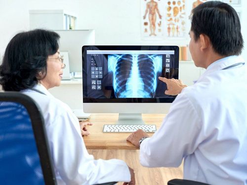
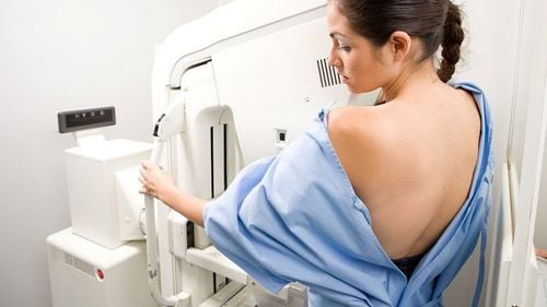
Chest X-ray patient receives about 0.1 mSv, equal to the amount of radiation that humans are exposed to from the natural environment in 10 days. Human mammograms received 0.4 mSv, equivalent to the effect of natural background radiation for 7 weeks. Some other imaging tests that have higher levels of radiation include:
Baryte colonoscopy: the patient received 8 mSv of radiation, equal to the expected natural human exposure in 3 years. CT scan (computed tomography) of the abdomen and pelvis: the person being scanned receives about 10 mSv. PET/CT scan: the patient receives about 25 mSv of radiation, which is equivalent to about 8 years of average background radiation exposure. However, the above are only general estimates for an adult of average body size. Studies have found that the actual amount of radiation each person receives can vary from the levels illustrated above.

Chụp X quang ngực giúp chẩn đoán ung thư phổi và các bệnh quan trọng khác với lượng phóng xạ rất thấp.
3. What to do when asked to conduct diagnostic imaging exams?
If you are concerned about the amount of radiation from the CT scanner, PET/CT scanner, or any other diagnostic imaging exam that uses radiation, make it clear to your hospital doctor about your concern. his mind. It is important to make sure that the examination is absolutely necessary and the best course of action to use in your case. Also, ask your doctor how beneficial the results of the test will be afterwards.
Many health professionals recommend that you only have diagnostic imaging tests when needed, trying to limit your exposure to all forms of radiation as little as possible. You must tell your healthcare provider right away if you are pregnant. In cases where some radiation exposure is required, ask your healthcare provider to cover other parts of your body that don't need testing. A lead apron can be used to protect your parts such as your chest or abdomen from radiation, a lead collar is a protective shield for the thyroid gland.
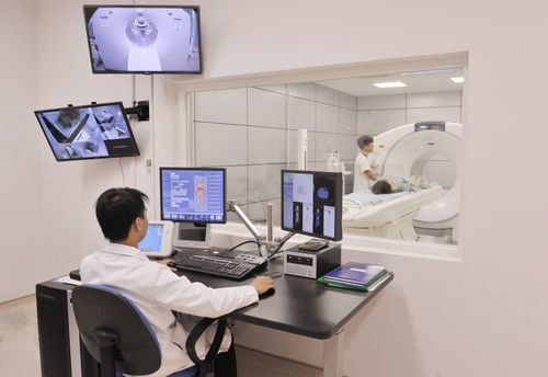
It is advisable to save the results of the scans to keep track of the examination history and to provide to other doctors in the future if available. This will save you from having to do the same tests again. Note that you can rest assured when you have MRI and ultrasound because you are not exposed to radiation.
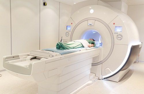
Chụp cộng hưởng từ MRI người bệnh sẽ không tiếp xúc được với bức xạ
4. Children and radiation from diagnostic imaging
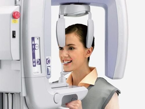
Professionals know that children are more sensitive to radiation than adults and need to be protected from radiation to the maximum extent. Therefore, doctors often focus on reducing radiation exposure for pediatric patients if they have to undergo radiation imaging tests. However, parents should ask the following questions before conducting the test to get a clear explanation from the medical staff:
Why does the child need imaging tests? What type of imaging test is appropriate for a child? Does that form of testing use radiation? Are there other ways to choose without using radiation? Can the amount of radiation used be adjusted to make it safer for young children? If the benefits of screening outweigh the risks of radiation exposure, parents should be reassured that their child is being tested. Parents also pay attention to save the results of the child's imaging tests to monitor and provide to doctors in other visits later.

Phụ huynh nên làm rõ các thông tin với bác sĩ trước khi để trẻ xét nghiệm có dùng phóng xạ
5. Cancer risk from radiation exposure
How radiation affects people depends on the following factors:
Type of examination performed Part of body area exposed Patient height and weight Age and sex Some another factor. According to medical experts, the increased risk of cancer by radiation from imaging tests is very small. On the other hand, it is difficult to determine how much radiation exposure from these examinations is associated with an increased risk of cancer in a person. Most studies on radiation risk and cancer only look at people exposed to very high doses of radiation, such as uranium miners and people affected by atomic bombs. If radiation exposure is low, it is difficult to calculate and draw conclusions.
A normally healthy person is still being exposed to radiation from all sources in nature with increasing levels towards the end of life. Furthermore, because radiation can increase the risk of cancer, imaging tests that use radiation are only performed when absolutely necessary with good reason. If possible, non-radiation imaging studies such as ultrasound or MRI should be selected. However, when an X-ray, CT scan or PET/CT is the best way to find the disease, the risk of radiation effects is negligible compared to the benefits of the examination results.
Please dial HOTLINE for more information or register for an appointment HERE. Download MyVinmec app to make appointments faster and to manage your bookings easily.
Reference source: Cancer.org