This is an automatically translated article.
The article was professionally consulted with Master, Doctor Tong Van Hoan - Resuscitation - Emergency Doctor - Emergency Department - Vinmec Danang International Hospital.1. Symptoms, how to diagnose pericardial effusion
Pericardial effusion is the accumulation of excess fluid in the pericardial cavity around the heart. The pericardium does not expand easily; As a result, fluid buildup rapidly leads to increased pressure around the heart. An increase in pressure restricts cardiac filling, leading to decreased cardiac output and cardiac tamponade.Signs and symptoms are common in cardiac tamponade and include dyspnea, hypotension, muffled heart sounds, coronary artery distension, and arrhythmias. Diagnosis of pericardial effusion by echocardiography. Small effusion in stable patient receiving medical treatment. Larger effusions and cardiac tamponade may require pericardiocentesis or pericardiocentesis.
1.1. Symptoms of pericardial effusion causing acute tamponade
Most of the physical symptoms are nonspecific and include:Tachycardia.
Hypotension (with or without shock) with orthostatic hypotension.
Hearing the heart can see faint heart sounds.
Symptoms like right heart failure: Enlarged liver, distended neck veins...
Signs of inversion is defined as a drop in blood pressure > 10 mmHg when the patient takes a deep breath.
Other physical signs include cold extremities, tachypnea, hepatomegaly, and signs of pericardial effusion.
In some severe conditions (left ventricular free wall rupture in acute MI, aortic rupture in aortic dissection, etc.) patients often come to the hospital with circulatory arrest (usually dissociation). electromyography) or hypotension, dizziness, confusion or shock.
Symptoms of the causative agent of pericardial effusion.
Severe progressive cardiac tamponade can lead to complications including liver failure, renal failure, mesenteric ischemia...
1.2. Diagnosis of pericardial effusion causing acute tamponade
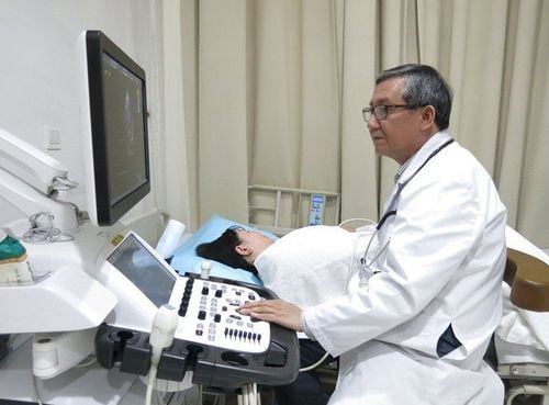
1.2.1 Transthoracic echocardiography:It is a mandatory method to perform when clinically suspected pericardial effusion (fluid in the pericardial cavity indicated by ultrasound gaps). Signs of tamponade include:
The right atrial compression sign, diastolic usually begins at the end of diastole and is most evident in the left parasternal view of the transverse, subcostal, and 4-chamber apex.
Right ventricular compression is usually observed in the anterior wall of the right ventricle and the funnel region in the supine position. The left sternal longitudinal and transverse axes are the two most favorable views to observe this sign. A TM ultrasound should be used to confirm this finding. Isolated right ventricular compression on echocardiography may precede the clinical presentation of tamponade.
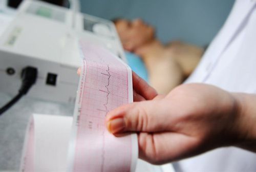
1.2.2 Electrocardiogram:
Low voltage of QRS complex: In peripheral leads total amplitude of QRS < 5 mm, in precordial leads total amplitude < 10 mm.
Alternating electrophysiological images: Low amplitude QRS complexes interspersed with high amplitude QRS complexes with sinus tachycardia.
1.2.3 Straight chest radiograph:
bulbous tachycardia + bright lung field (if there is previous pericardial effusion).
Cardiac shadow may be normal (if there is no previous pericardial effusion.
Note: fluid < 250 ml usually does not enlarge the heart but can cause fatal acute cardiac tamponade.
1.3. Diagnosis
The diagnosis of pericardial effusion with tamponade is when one of the following two criteria is met:
Inversion pulse: systolic blood pressure decreases > 10 mm Hg during inspiration while diastolic pressure remains unchanged
Upper cardiac wall compression echocardiogram (atrial compression or ventricular compression).

3. Relationship of pericardial effusion and acute cardiac tamponade
Cardiac tamponade is a life-threatening slow or rapid cardiac compression caused by increased pericardial fluid. The pericardium can stretch to accommodate the accumulation of fluid, but when it reaches a certain limit the pericardium cannot increase any more, an equalization of pressure occurs in the pericardium (central veins, right heart chambers) and left), usually around 15-20 mmHg. At this point, the right ventricle collapses and the hypotension becomes severe.The hemodynamic effects of a significant pericardial effusion are a continuum because the small increase in pericardial content joins the pericardium to the heart, significantly increasing the atria and especially the ventricles interactions. . Such phenomena increase the normal reciprocal respiratory effects on the right and left sides of the heart.
Rate of fluid accumulation is also important: If fluid enters the pericardial cavity rapidly (eg, chest trauma), as little as 150 mL can lead to compression. If fluid builds up slowly, the pericardial sac may stretch to hold about 2 L of fluid. During the first symptoms of hemodynamic deterioration of a pericardial effusion, the patient may only find echocardiography to detect chamber compression
4. Treatment of pericardial effusion causing acute tamponade
4.1 General principles Once a chest compression has been diagnosed, the first priority should be to drain the pericardial fluid.The method of implementation can be percutaneous aspiration with local anesthesia, surgical drainage (opening the pericardial cavity under the bone, sternum). In the case of postoperative TMD, surgical drainage of the pericardium should be indicated.
4.2 Indication for pericardiocentesis: In hemodynamically unstable patients, emergency procedure is required because only fluid removal can normalize ventricular filling and restore full cardiac output. enough. Otherwise, the procedure can be done within a few hours of the exam and the most appropriate instructional and visual approach can be planned. The indication for urgent puncture or drainage in patients with suspected compression depends on: etiology, clinical presentation, and echocardiographic findings.
In the case of pericardial effusion without hemodynamic compromise, pericardiocentesis is indicated for moderate to large symptomatic pericardial effusion unresponsive to medical treatment, or in cases of pericardial effusion. Smaller pericardial effusion, when tuberculosis, bacterial, or cancerous pericarditis is suspected, or in chronic cases (lasting more than three months), large pericardial effusion (> 20 mm on echocardiography at heart rate). stretch).
Pericardial puncture for diagnostic purposes has not been demonstrated in cases of mild or moderate effusion (<20 mm) for the following reasons:
Low diagnostic potential (the underlying pathology is often established). known or determined through various noninvasive tests). Viral (idiopathic) pericarditis is usually self-limited and requires only anti-inflammatory therapy. The surgical risk is high compared to the low diagnostic outcome.

However, if immediate surgical management is not available, or if the patient is very unstable, pericardiocentesis can be attempted and a very small amount of blood drained to maintain blood pressure at approximately 90 mmHg as a bridge for emergency surgery. Relative contraindications include uncorrected coagulopathy, anticoagulant therapy, and thrombocytopenia (PLTc <50,000/mm3).
4.4 Procedure Procedure Removal of pericardial fluid, with or without echocardiographic guidance, is definitive therapy for compression and can be accomplished by the following 3 methods.
4.4.1 Emergency percutaneous drainage: This is a life-saving bed procedure. Epidural approach. Therefore, pericardiocentesis performed without echocardiographic guidance is safest. A G16 or G18 needle is inserted at an angle of 30-45° to the skin, near the left xiphocostal angle, toward the left shoulder. When performed as an emergency, this procedure is associated with a reported mortality rate of approximately 4% and a complication rate of 17%.
4.4.2 Video-Assisted Thoracic Surgery (VATS) Also known as thoracoscopy is a minimally invasive technique performed under general anesthesia. VATS allows visual assessment of the pericardium and is used when a pericardial effusion has not been diagnosed despite the results of previous less invasive tests. It is also used to drain excess fluid and prevent its accumulation.
4.4.3 Echocardiogram-guided pericardiocentesis This procedure is usually done in the ultrasound room. The location is usually determined in the left intercostal space. First, mark the site of penetration based on the area of maximum fluid accumulation closest to the transducer. Then, measure the distance from the skin to the pericardial cavity. The angle of the probe should be the trajectory of the needle during the execution. Avoid the inferior edge of the rib during needle insertion to avoid nerve damage. Leave a size 16 catheter in place for continuous drainage.
4.4.4 Percutaneous balloon pericardiocentesis A nonsurgical procedure performed using X-ray guidance to view the pericardium and insert a balloon dilatation catheter. This trick is not common.
4.4 Complications of pericardiocentesis
The rate of major complications reported in large observational studies for echo-guided pericardiocentesis or fluoroscopy was 0.3-3.9%, and the rate of minor complications was is 0.4-20%.The most serious complications include:
Death Heart chamber damage Coronary or intercostal artery tear Perforation of abdominal viscera or peritoneal cavity Pneumothorax requiring chest tube Pneumopericardial arrhythmia Ventricular arrhythmia and syndrome decrease in pericardial pressure Myocardial and coronary perforations may initially be silent and occur after hemothorax or bradycardia in bradycardia. Pericardial hypotension is a rare, potentially life-threatening syndrome characterized by broad clinical scenarios (ranging from pulmonary edema to cardiogenic shock). Acute left ventricular overload due to right anterior preload associated with persistent catecholaminergic peripheral vasoconstriction. To date, there are no effective recommendations for the prevention of this syndrome except to remove enough fluid to normalize central venous and systemic blood pressure (no more than 1L) and complete elimination in next few hours. Minor complications include:
Transient vascular hypotension Bradycardia, supraventricular arrhythmias Pneumothorax without hemodynamic instability Pleural fistula.
4.5 Tracking
MNT fluid analysis: cell count, culture, cytology to determine etiology (malignancy) and long-term treatment.Before removing the catheter, echocardiography should be done to check the MNT fluid.
Take a chest X-ray immediately after aspiration to rule out pneumothorax.
Why should you choose Vinmec?
Emergency and Resuscitation Department of Vinmec medical system with a team of experts, experienced in emergency practice of cardiovascular diseases, in addition, the system is also fully equipped with modern equipment and machinery. It is a prestigious and reliable first aid facility for patients.
Please dial HOTLINE for more information or register for an appointment HERE. Download MyVinmec app to make appointments faster and to manage your bookings easily.
References: my.clevelandclinic.org, amboss.com, escardio.org, emedicine.medscape.com, lecturio.com


