This is an automatically translated article.
Article written by Doctor Han Thu Huong - Vinmec High-Tech Center
Chromosomes are DNA-packing structures found inside the body's cells. Humans have 23 pairs of chromosomes (46 chromosomes in total). Each copy in a pair comes from the mother (egg), the other comes from the father (sperm).
1. Chromosomes
The first 22 pairs are called autosomes and are the same in both males and females. The 23rd pair of chromosomes is called the sex chromosome (X and Y). Females have two copies of the X chromosome, one X from their father and one X from their mother. Males have one X chromosome from their mother and one Y chromosome from their father. Mothers always pass on the X chromosome (to their son or daughter) while fathers can either contribute X or Y, determining the sex of the child.
Hình 1. Karyotype bình thường ở nam giới và nữ giới (Cytogenetics Gallery)
2. Chromosomal Abnormalities
Chromosomal abnormalities are divided into 2 main types: quantity disorders and structural disorders. Most abnormalities appear in the sex cells of the parent's body, but there are also cases caused by genetics from previous generations.
2.1. An aneuploidy is the result of failure to segregate during meiosis, in which pairs of homologous chromosomes move to the same daughter cell. So the fertilized egg will receive one or three copies of the chromosome instead of the usual two. Because they involve many genes, altering the normal genome balance, most chromosome number disorders cause embryonic death, especially chromosome loss. Non-lethal disorders often lead to infertility, because they interfere with normal cell division.
The best known and most common chromosomal abnormality is Down syndrome (trisomy 21), in addition to some other common disorders of chromosome number such as trisomy 13, trisomy 18, Klinefelter syndrome and Turner syndrome...
2.1.1. Autosomal-associated disease Down syndrome Down syndrome (3 chromosomes 21 or trisomy 21) is the most common chromosomal disease of live neonates, occurring in approximately 1 in 660 live births. The risk of having a baby with Down syndrome increases with maternal age, so women over 35 should be fully screened, especially during pregnancy.
Common features: developmental delay and mild to moderate intellectual disability; typical facial features: small head, flat face, protruding tongue, slanted eyes, flat nose, short neck; structural heart defects, malformations of the gastrointestinal tract, duodenal stenosis, dilated colon; low or poor muscle tone; can live to adulthood.
Down's disease is not treatable but can be diagnosed early. Comorbidities such as congenital heart defects can be treated with surgery. Understanding the disease and implementing early intervention will make the life of people with Down syndrome better.
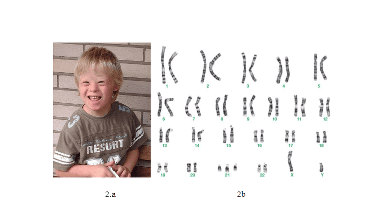
Hình 2.a: Cậu bé 8 tuổi mắc hội chứng Down (Wikipedia) Hình 2.b: Karyotype của nam giới mắc hội chứng Down (Karyotype Analyses of Down Syndrome Children in East Priangan Indonesia)
Edward syndrome (3 chromosomes 18 or trisomy 18) occurs in about 1/3333 live births. Children with this syndrome can live no more than 1 year old. Common features: round face, small head, small chin; malformations of the heart, kidneys and some other organs; increased muscle tone; hand and/or foot abnormalities; developmental delay and severe intellectual disability. Patau syndrome (3 chromosomes 13 or trisomy 13) occurs in about 1 in 5000 live births. Children with this syndrome can live no more than 1 year old. Common features: typical face with cleft lip and jaw, small eyes, extra little finger; heart, brain, kidney abnormalities; severe intellectual and developmental disabilities. 2.1.2. Sex-linked pathology Turner syndrome (1 X chromosome or monosomy X) occurs in about 1 in 2,000 live births. Common features: short, well-proportioned; structural defects of the heart; Primary ovarian dysfunction leads to amenorrhea and infertility. Triple X syndrome (3 X chromosomes) occurs in about 1 in 1000 female live births. Many women with this syndrome often have no outward symptoms. Common features: above average; learning difficulties, slow language development; slow development of motor skills; behavioral and emotional difficulties; normal sexual development and fertility. Klinefelter syndrome (47,XXY) occurs in about 1 in 1000 male live births. Common features: above average height, disproportionately long arms and legs; underdeveloped testicles, infertility; Difficulty learning, slow language development. Jacobs syndrome (47,XYY) occurs in about 1 in 1000 live births. Common features: learning difficulties, slow language development; increased risk of ADHD and autism spectrum disorders; normal fertility.
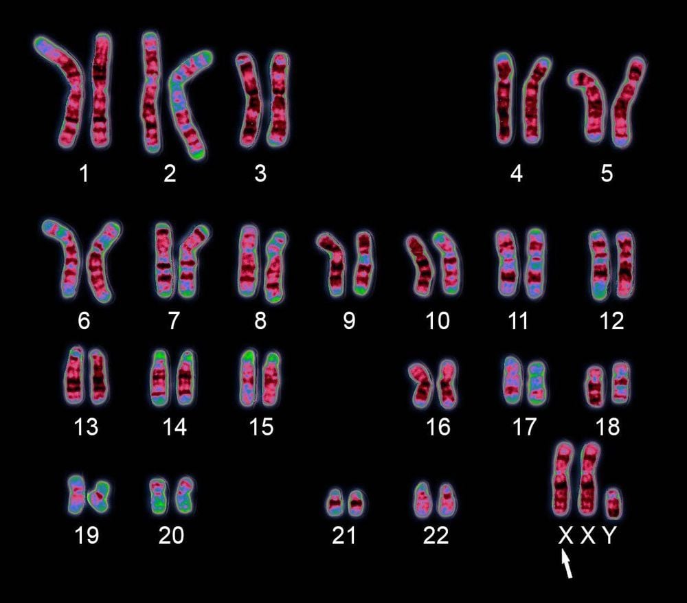
Hội chứng Klinefelter (47,XXY)
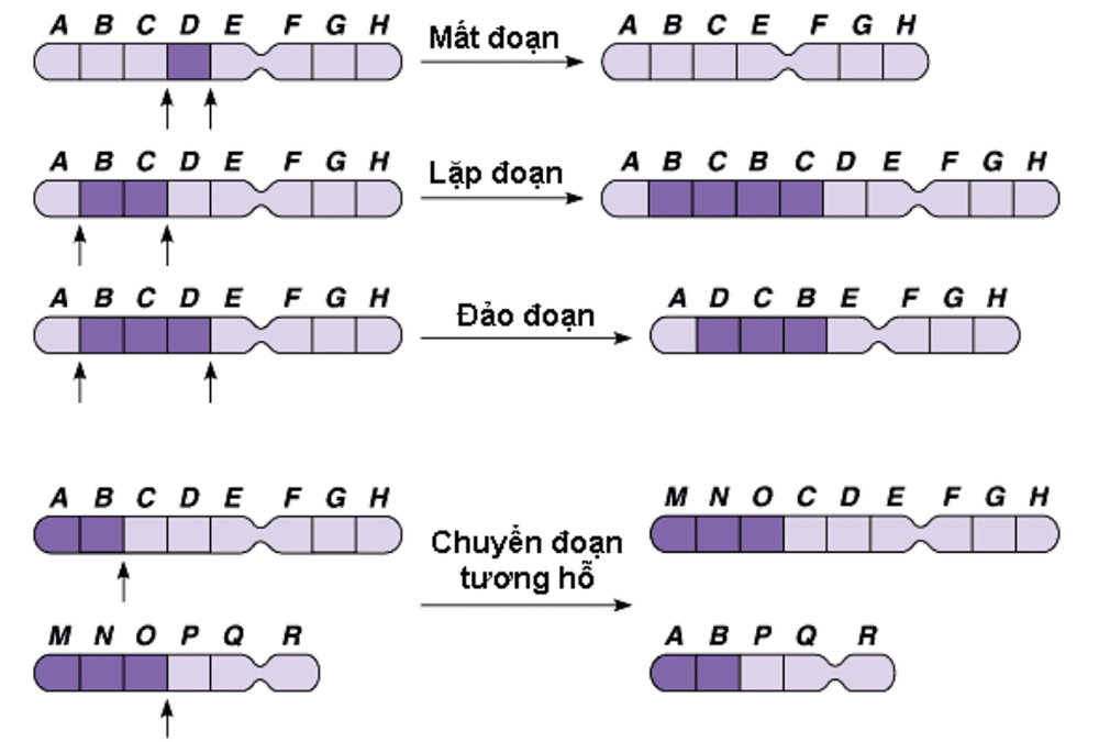
2.2. Chromosome Structural Abnormalities Chromosome structural abnormalities are the result of incorrect breakage and splicing of chromosome segments. A variety of structural chromosomal abnormalities lead to pathology.
2.2.1. Chromosome deletion The term "deletion" simply means a part of a chromosome is missing or "lost". Deletions can occur in any part of any chromosome. A very small number of deletions are called micro-deletions. Deletions and microdeletions can lead to intellectual and developmental disabilities as well as birth defects. Some common deletion diseases:
Cri du Chat syndrome (cat syndrome), is one of the rare genetic diseases caused by the loss of the short arm of chromosome number 5. The incidence is 1/ 15000-1/50000 live births. Babies born with this syndrome often have small heads, low birth weight, muscle weakness, mental retardation, and difficulty in eating and breathing. In particular, the baby's cry resembles a cat's meow. Wolf-Hirschhorn syndrome (4p syndrome), caused by a 1p36 deletion on chromosome 4, has an incidence of approximately 1 in 50,000 live births. Signs and symptoms include growth deficiency, hypotonia, craniofacial features, intellectual disability, and cardiac and brain abnormalities. Average life expectancy depends on the severity of the features. DiGeorge syndrome (deletion syndrome 22q11.2) is a syndrome caused by microdeletions in the long arm of chromosome 22, affecting about 1 in 4000-1/6000 live births. Digeorge syndrome can cause many different problems that affect many different parts and parts of the body. 5 common signs of DiGeorge syndrome: congenital heart defects; characteristic facial features (low ears, wide eyes, small jaw, narrow groove on the upper lip); cleft palate ; low blood calcium levels (causing seizures) due to hypoparathyroidism; often infections due to immunodeficiency, or autoimmune disease (Basedow's disease, chronic arthritis). Prader-Willi syndrome (deletion syndrome 15q11-q13), caused by a deletion on the long arm of chromosome 15, occurs in approximately 1/10,000-25,000 live births. Prader willi syndrome affects people's physical, mental, linguistic and behavioral health. More specifically, the disease also disrupts the ability to eat, making children feel unfulfilled. So common complications of this disease are obesity and more serious diseases such as type 2 diabetes, cardiovascular disease and stroke, arthritis, sleep apnea, hypogonadism, infertility. or osteoporosis... Life expectancy is normal, but may decrease depending on severity of symptoms.

Hội chứng Prader-Willi gây biến chứng béo phì
2.2.2. Chromosome duplication Duplication, sometimes known as partial trisomy, occurs when there is an extra copy of a chromosome segment. Like deletions, duplications can occur on any part of the chromosome. Chromosome duplication can lead to intellectual disabilities as well as birth defects.
Pallister Killian syndrome is the result of the short-winged duplication of chromosome 12. The disease is usually mosaic, in the patient's body there are 2 cell lines, 1 cell line has an extra short arm of chromosomes 12 and 1. normal line (46 chromosomes and no extra genetic material). Common manifestations include severe intellectual disability, poor muscle tone, "rough" facial features and prominent forehead, very thin upper lip, thicker lower lip, and short nose. Other health problems include stiff joints, cataracts in adulthood, hearing loss, and heart defects. People with Pallister Killian syndrome have a short life expectancy, but can live to the age of 40. 2.2.3. Chromosome translocation Chromosomal translocation is a phenomenon of exchanging segments between two chromosomes, there are two types of translocations: reciprocal translocations and concordant translocations.
Mutual translocation: each chromosome breaks in one place and exchanges the break, forming two new chromosomes and both change morphology if the swap segment is different in size. Reciprocal translocation carriers do not lose genetic material and have normal phenotypes, so they are called balanced translocations. Balanced reciprocal transitions appear at a scale of 1/500.
The most common translocation is chronic myelogenous leukemia (CML), a common blood cancer. Part of chromosome 9 translocates with part of chromosome 22, creating the Philadelphia chromosome. The Philadelphia chromosome is present in the blood cells of 90% of people with chronic myeloid leukemia.
Concordant transposition (Robertson translocation): reciprocal translocation and occurs only with the centromere chromosome, in which two chromosomes are broken across the proximal region, the breaks switch each other to form an abnormal chromosome contains the long arm of the two transpositioned centromeres and a very small chromosome containing the short arm of the two transpositioned centromeres, which will be destroyed. The rate of concordant translocation is 1/1000 people in the population.
Carriers of balanced reciprocal and concordant transposition often have no clinical manifestations but may be at risk: infertility, recurrent miscarriages, birth defects, birth defects, intellectual disabilities and developmental disabilities.
2.2.4. Chromosome inversion Inversion occurs when one chromosome is broken in two regions and the resulting piece of DNA is reversed and inserted back into the chromosome. Although there are possible effects on fertility due to unbalanced chromosomes due to crossovers in the inversion region in heterozygotes, for some cases there is no risk of miscarriage or problems. on chromosome segregation during cell division.
However, not all inversions are harmless, some pathologies are sometimes found as a result of inversions, mainly due to direct disruption of a gene or to altered gene expression. These mutations are present in patients either as genetic mutations confined to a given family, and therefore they do not represent segregated polymorphic variants in the human population. However, they are of clinical importance and may contribute to the identification of genes underlying some rare disorders.
2.2.5. Ring chromosomes Ring chromosomes form when the ends of two arms of the same chromosome are broken, which then fuse together to form a ring-shaped chromosome. Deletions at the ends of both arms of the chromosome lead to a lack of DNA, which can cause chromosomal disorders. The typical pathology for this abnormality is chromosomal 14 syndrome.
14 chromosomal syndrome is a disease characterized by epileptic status and intellectual disability. Other features of ring 14 syndrome may include slow growth and short stature, small head (microcephaly), puffy hands and/or feet due to fluid accumulation (lymphedema), and some barely noticeable difference on the face.
3. Test for chromosomal pathology
Suspicion of chromosomal abnormalities in some cases:
Before birth: pregnant women are prescribed amniocentesis or placental biopsy to analyze fetal chromosomes in some cases such as pregnant women over 35 years old, have money History of spontaneous abortion, stillbirth, history of birth defects, abnormal fetal ultrasound results, high-risk serological screening results, pregnant couples One of the two women has been identified with a genetic structural mutation... After birth: newborn with congenital malformations, unexplained psychomotor retardation, family history of people with chromosomal mutations, primary or secondary infertility, primary or secondary amenorrhea, couples with consecutive miscarriages or stillbirths, blood cancers, etc. Certain types of genetic testing can be performed. identification of chromosomal disorders:
Karyotyping (Chromosome Formula) FISH (Fluorescent In situ hybridization) Array-CGH (Comparative Genome Hybridization)

Xét nghiệm di truyền trước mang thai là việc làm cần thiết
3.1. Karyotyping Karyotyping is a technique for testing the number and structure of human chromosomes based on cell culture. The test result is a karyotype with the chromosomes sorted and numbered by size from largest to smallest.
With G-band staining technique, the resolution of chromosomes is usually in the range of 350-850 bands on haploid sets with each band size from 5-10Mb at normal resolution and each band size from 3- 5 Mb at high resolution, so this technique only allows detecting anomalies larger than 3 Mb. Cases of chromosomal abnormalities smaller than the above size will not be detected.
3.2. FISH (Fluorescent insitu hybridization) FISH is a technique that mediates between cellular and molecular genetics, using a specific primer (probe) attached to a fluorescent signal, to detect the presence of fluorescence. presence or absence of a certain gene segment on the chromosome, with the advantage of fast, specific and accurate.
FISH can detect small deletions that are difficult to detect on conventional chromosomal formulas, some specific translocations such as translocations of chromosomes 9 and 22 (Ph1), 4 and 11, 8 and 21... have classification and prognostic value in cancer treatment, or used in prenatal diagnosis of chromosomal abnormalities.
3.3. Array-CGH (Comparative Genome Hybrid) Array CGH is a technique to compare the DNA sample to be analyzed with the control DNA sample to detect abnormalities on chromosomes with a resolution of 100kb-1Mb, so array-CGH is often considered is molecular karyotyping.
Chromosomal abnormalities that can be identified by array CGH: aneuploidies and mosaic cases, types of unbalanced translocations, marker chromosomes, syndromes associated with microdeletions, microdelegation of chromosomes that Karyotype and FISH could not detect, cases of unbalanced rearrangement of the paraterminal region of chromosomes with high accuracy and short duration. However, this technique cannot detect balanced rearrangements of chromosomes such as balanced translocations, inversions, Robertson translocations, reciprocal insertions, point mutations, and mosaicism. short.
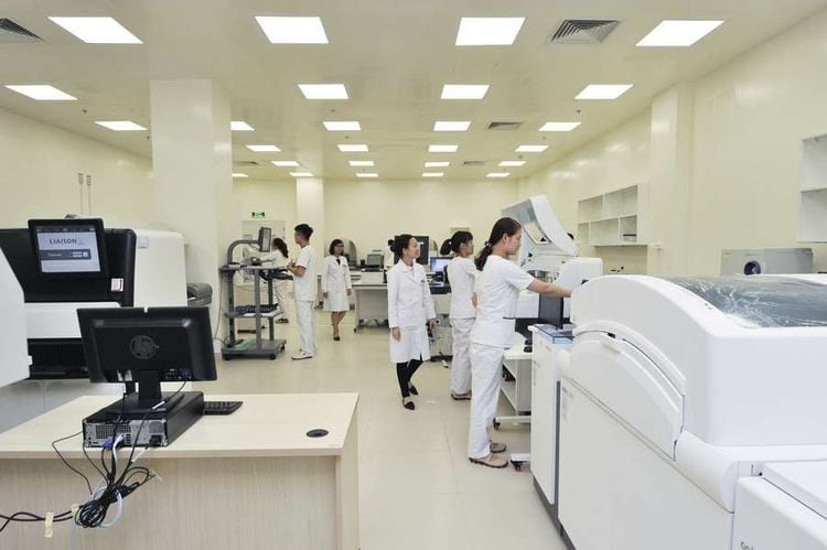
Hệ thống labo xét nghiệm tiêu chuẩn quốc tế tại Vinmec
Vinmec International General Hospital is one of the hospitals that not only ensures professional quality with a team of leading medical doctors, modern equipment and technology, but also stands out for its examination and consultation services. comprehensive and professional medical consultation and treatment; civilized, polite, safe and sterile medical examination and treatment space.
Customers can directly go to Vinmec Health system nationwide to visit or contact the hotline here for support.
References
Chang-Hui Shen. Chapter 13: Molecular Diagnosis of Chromosomal Disorders . Diagnostic Molecular Biology, 2019. Fred Levine. Basic Genetic Principles . Fetal and Neonatal Physiology (Fifth Edition), 2017. Hindi E. Stohl, Lawrence D. Platt. Introduction to Aneuploidy . Obstetric Imaging: Fetal Diagnosis and Care (Second Edition), 2018. Mary E. Norton, Jeffrey A. Kuller and Lorraine Dugoff. Chapter 4 - Cytogenetics: Part 1, General Concepts and Aneuploid Conditions . Perinatal Genetics, 2019. Mary E. Norton, Jeffrey A. Kuller and Lorraine Dugoff. Chapter 5 - Cytogenetics: Part 2, Structural Chromosome Rearrangements and Reproductive Impact . Perinatal Genetics, 2019. Sameer M. Zuberi. Pediatric Neurology Part I . Handbook of Clinical Neurology, 2013. MORE:
Role of chromosomal testing in the diagnosis of fetal malformations Chromosome testing in the diagnosis of infertility Misinformation about X and Y sperm


