This is an automatically translated article.
The article is professionally consulted by Master, Doctor Tong Diu Huong - Radiologist - Department of Diagnostic Imaging - Vinmec Nha Trang International General Hospital.The method of placing chemotherapy under the skin is used to deliver chemotherapy to cancer patients. With the help of digital imaging, the procedure is improved in accuracy, safety, and reduces complications for the patient.
1. What is a subcutaneous chemical infusion chamber?
Subcutaneous chemotherapy (chemoport) is a medical device that is used to deliver chemotherapy to cancer patients with indications for chemotherapy.Chemical infusion chamber is a system consisting of a catheter (catheter) and injection chamber (portal chamber). The catheter part is placed into the large vein system (including external jugular vein, subclavian vein, brachial vein ..), while the injection chamber is completely implanted into the subcutaneous tissue of the patient.
In the past, the subcutaneous chemical infusion chamber was often placed quite subjectively, based on the experience of the operator and had many risks leading to serious complications, such as:
Pneumothorax; Tear of the subclavian artery; Arteriovenous catheterization; Missing the catheter. The application of digital imaging to erase the background to place the chemical infusion chamber will have the advantage of improving accuracy, increasing safety and reducing significant complications for patients.

Buồng truyền hóa chất dưới da trước đây có thể gây tràn khí màng phổi
2. When is the need for subcutaneous chemotherapy?
Subcutaneous chemotherapy is often indicated in the following specific cases:Cancer patients need to receive consecutive chemotherapy infusions and chemotherapy drugs can cause vein damage or blood clots; Patients who are having problems related to the digestive system or have a poor prognosis, need to supplement with combined nutrition in the postoperative period and long after undergoing chemotherapy and radiation therapy; Cancer disease has been treated stably, it is expected to use chemotherapy chambers for palliative care and supportive pain treatment in cases with a prognosis of more than 3 months; Need for drug infusion into the central vein, blood transfusion, long-term fluid infusion for the patient over a period of 3 to 10 years; Children or adults requiring complete parenteral nutrition; Effective support for nurses and family doctors in home care.
3. Where in the body is the subcutaneous injection chamber implanted?
To perform subcutaneous chemotherapy, the patient needs to undergo a relatively simple procedure. Accordingly, the subcutaneous injection chamber is completely implanted inside the body, one side of the catheter is inserted into the vein, the other side is connected to the injection chamber and completely implanted under the skin to deliver chemicals into the muscle. body. The most common location for the infusion chamber is subcutaneously in the chest wall and into the subclavian and superior vena cava.The facility that places the subcutaneous injection chamber usually issues a card to the patient to note specific information. The skin where the injection chamber is implanted will be raised, similar to a coin inserted under the skin, and the patient will also easily adapt to this feeling after a short time. The subcutaneous chemical infusion chamber is normally implanted in a discreet location, without affecting the patient's aesthetics and daily activities.
The life of the injection chamber depends on the type, care, adaptation of the body and the number of times the needle is pierced through the membrane of the injection chamber (average 1000 - 3600 times). However, patients should contact the hospital where they placed the subcutaneous chemotherapy chamber for an accurate answer, in accordance with the current situation.
4. The procedure of placing the chemical infusion chamber under the skin under the digital scan to erase the background
4.1. Open access to the central vein
Usually, puncture of the internal jugular vein - right (position of supraclavicular fossa). In addition, it is also possible to puncture the subclavian vein depending on the case; Local anesthetic, then skin incision; Use an 18G needle to puncture the vein under ultrasound guidance; Under digital subtraction angiography (DSA), insert a lead and a 5F dilatation tube into the superior vena cava.4.2. Place an infusion chamber under the skin of the chest wall
Extensive local anesthesia in the skin and subcutaneous tissues of the chest wall, as well as the subclavian region; Incision of the skin and subcutaneous tissue of the chest wall, making it wide enough to place the infusion chamber.4.3. Place the tube straight into the superior vena cava
Create a subcutaneous tunnel, from the portal site to the venous puncture site in the right supraclavicular fossa; Under fluoroscopic guidance, measure the catheter just enough to reach the junction of the superior vena cava and the right atrium; Cut off the excess cannula; Insert the catheter through the tunnel just created to reach the site of internal jugular vein puncture; Under the guidance of digital background erasure, the catheter is inserted into the superior vena cava through the lead.4.4. End
Suture the skin at the site of the subclavian vein puncture and the placement of the subcutaneous chemotherapy chamber; Check the circulation of the infusion chamber and catheter.5. Evaluation of results
The medial end of the chemotherapy infusion chamber is positioned where the superior vena cava empties into the right atrium; The outer end of the subcutaneous chemotherapy chamber is located properly in the subcutaneous soft part of the chest wall; Take the syringe and insert it into the infusion chamber, suck it out, and see blood.
Đặt buồng truyền hóa chất dưới da dưới chụp số hóa xóa nền có thể gây nhiễm khuẩn máu
6. Risk of complications and treatment
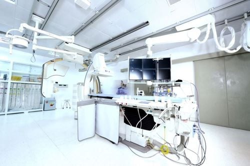
Pneumothorax: Bandage the drainage site, with minimal pleural opening and continuous suction (if indicated); Hematoma under the skin of the chest wall: Apply pressure bandage to stop bleeding; Catheter rupture: Use a mesh trap to remove the broken catheter or perform surgery if the technique fails; Bacterial infections under the skin of the chest wall, around the port or along the internal jugular vein: Cleaning combined with topical and systemic antibiotics; Blood infection : Need specialist consultation. In general, the procedure for placing a chemical infusion chamber under the skin is relatively simple, but still requires an experienced physician. The subcutaneous chemotherapy chamber will be implanted completely inside the body, one end of the catheter is inserted into a vein, the other end connects to the implantation chamber in front of the chest or in front of the arm. Patients and relatives need to know how to take care of the subcutaneous chemotherapy chamber, as well as immediately notify the doctor if any problems are detected.Vinmec International General Hospital has applied digital imaging technology to erase the background in examination and diagnosis of many diseases. Accordingly, the dsa imaging technique at Vinmec is carried out methodically and according to standard procedures by a team of highly qualified medical professionals and modern machinery. Thereby giving accurate results, contributing significantly to the identification of the disease and the stage of the disease and timely treatment.
Master. Doctor. Tong Diu Huong has more than 10 years of experience in the field of specialized imaging in diseases on images Ultrasound, X-ray, multi-slice CT, Magnetic resonance in diseases of the nervous system, digestive system , urology, cardiology, musculoskeletal... Besides, there are interventional imaging techniques, fine needle aspiration cells under the guidance of ultrasound and computed tomography.
If you have a need for medical examination by modern and highly effective methods at Vinmec, please register here


