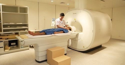
This is an automatically translated article.
The article is professionally consulted by Dr. Nguyen Dinh Hung - Diagnostic Imaging - Department of Diagnostic Imaging - Vinmec Hai Phong International General Hospital.
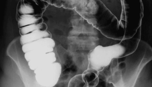
Digital X-ray of the colon is a technique to contrast the colonic frame with Baryt's suspension. The technique is performed in the condition that the patient is completely enema removed before the contrast agent is inserted.
1. Preparing for a digital colon x-ray
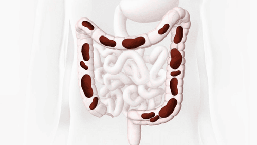
Digital X-ray technique of colon is indicated to investigate diseases of the colon. For patients with symptoms of colitis such as abdominal pain, prolonged digestive disorders, bloating, gas, change in stool pattern, loss of appetite, fatigue, rapid weight loss will be prescribed by a doctor. colon x-ray.
The technique has no absolute contraindications, but it should be noted with pregnant women. Colon x-ray is performed by a specialist, an electro-optical technician with a full range of facilities including x-ray machine, film, film printer, and normal working storage system.
In addition, the consumables required in the process of digitizing colon x-ray are:
● Barytite contrast agents diluted 30-40%
● Water-soluble iodine contrast agents are used. for abdominal emergencies because it can be drained in a few hours, but because of its poor adhesion, hypertonicity, high cost, it is rarely used
Drugs to increase and decrease colonic motility
● Drugs to increase colon peristalsis
● Gloves, enemas
Patients need to have a diet that does not cause backlog before 2 days, and do not eat foods high in fiber or fermentation. Simultaneously use laxatives for 2 days and enema of the colon with 1.5-2 liters of warm water is given slowly at a height of 40cm, kept for 10 minutes, done 2 times a few hours apart or after 12 hours. before shooting.
2. Conduct digitized x-ray of colon
Injecting medicine
Take a non-standard abdominal film in the supine position, prepare a warm barytse placed 40cm above the table, insert the branched cannula into the anus. For Baryt to be gradually introduced, it is necessary to monitor under the television light to find the right position, minimize the radiographs, and reduce the radiation dose for both the doctor and the patient.
Perform the scan
The three-phase films are as follows:
Pre-medication to assess colon tone
● Take the drug after defecation to see the mucosa
● Inflate to create double contrast, see mucosal wall of colon
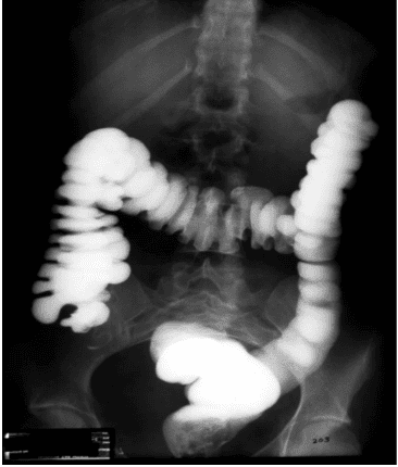
● Take the following postures in turn for evaluation:
Rectal oblique, posterior left, lateral: film 24x30cm
⮚ Sigma posterior left, oblique: film 24x30cm
Left splenic angle posterior right: 24x30cm film
⮚ Left posterior oblique angle: film 24x30cm
Caecum, left lateral ascending colon: film 24x30cm
⮚ Full film: 30x40cm film
⮚ With medication: 30x40cm film
⮚ Inflatable: 30x40cm film
● Colectomy loop sigm-rectal position has the following positions:
⮚ Le Canuet position: two times, the left side and the ball
⮚ Chasard – Lapine position: the patient sits in the corner of the table, the body is bent, the center ray is residence L5.
3. Evaluation of the results
A clear digital x-ray image of the colon, fully showing the anatomical structures of the colonic framework, clearly showing the lesions if any. The doctor reads the lesion, describes it on a computer with an internal connection, prints the results, and can provide professional advice to the patient or family if required.

Gastrointestinal X-ray with contrast is an imaging method to evaluate the anatomical structure of the gastrointestinal tract. Therefore, patients should choose reputable medical facilities that have enough modern medical equipment systems to perform this technique.
In order to improve the medical examination and treatment process, Vinmec International General Hospital has now applied gastrointestinal X-ray technique in examination and diagnosis of many diseases. At the disease, Vinmec hospital system is built on the spirit of PATIENT CENTER. The equipment and machinery are invested in modern. The team of doctors with high professional qualifications, dedication to the profession, technical processes are elaborately built up to international standards. The examination and treatment will bring the patient the best results.
Doctor Nguyen Dinh Hung has over 10 years of experience in the field of diagnostic imaging (Ultrasound, CT, MRI). Trained and practiced on hepatobiliary interventional radiology at Bach Mai Hospital (Intervention under ultrasound guidance, DSA, CT...) and deployed at the Diagnostic Imaging Department of Viet Tiep Hospital Hai Phong. Currently, he is a doctor at the Diagnostic Imaging Department of Vinmec Hai Phong International General Hospital.
Please dial HOTLINE for more information or register for an appointment HERE. Download MyVinmec app to make appointments faster and to manage your bookings easily.


