This is an automatically translated article.
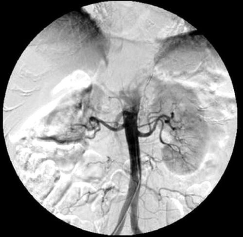
The article is expertly consulted by Master, Resident, Specialist I Trinh Le Hong Minh - Radiologist - Radiology Department - Vinmec Central Park International General Hospital.Digital angiography of the renal artery was performed using iodinated contrast agents to visualize the renal vasculature. When the capillaries and parenchyma are infiltrated, the renal morphology as well as the cortical medulla and calyx of the kidney are revealed.
1. Indications and contraindications for digitization of renal artery background
1.1 Designation
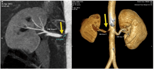
Blood in urine Suspicion of renal vascular disease: Renal stenosis, renal vascular malformation, aneurysm ... Angiography to serve for interventional radiology. Renal angiography in preparation for a kidney transplant. Evaluation of blood supply of kidney tumor diseases: Vascular lipoma... Renal trauma diseases with suspected vascular injury. Chronic inflammatory diseases with vascular damage.1.2 Contraindications
With digitized imaging of the renal artery, there is no absolute contraindication.However, digitization of the renal artery background is still relatively contraindicated in cases of coagulopathy, renal failure, a history of obvious allergy to iodinated contrast agents or pregnant women.
2. Preparing to conduct digital scan to erase the renal artery background
2.1 Digital angiography of renal artery background is performed by whom?
Specialist. Auxiliary physician. Nursing. Optical technician. Doctor, anesthesiologist (if the patient cannot cooperate).2.2 The equipment needed to prepare for digitizing the renal artery background
Digital background removal angiography (DSA). Dedicated electric pump. Film, film printer, image storage system. Lead vest, apron, X-ray shield.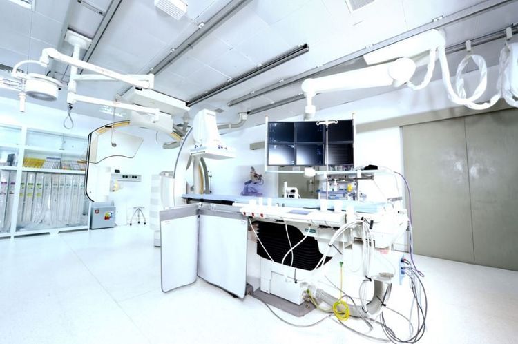
2.3 Common medical supplies to prepare
Syringe 1; 3; 5; 10 and 20 ml. Syringe pump for electric pump. Gloves, shirt, hat, surgical mask. Distilled water or physiological saline. Cotton, gauze, surgical tape. Medicine box and first aid kit for contrast drug accidents. Aseptic intervention kit: knife, scissors, tongs, 4 metal bowls, bean tray, tool tray.2.4 Special medical supplies used in digital imaging of renal artery background
Arterial puncture needle. The set of the internal circuit size 5-6F. Y-connector set. Three-pronged lock. Microcatheter 2-3F when ultra-selective imaging is required. Micro-conductor 0.014-0.018inch. Standard conductor 0.035inch. Angiography catheter size 4-5F. Instrumentation kit.2.5 Drugs used in digital angiography of renal artery background
Local anesthetic. Anticoagulants. Anticoagulant neutralizer. Iodine contrast agent is water soluble. Antiseptic solution for skin, mucous membranes, pre-anesthesia and general anesthesia (if there is an indication for anesthesia).2.6 Patients need to prepare before digitizing the renal artery background
Before conducting digital scan to erase the renal artery background, the patient must fast for 6 hours. However, the patient can still drink water but with less than 50ml of water.When entering the intervention room, the patient lies on his back and then installs the monitors to monitor breathing, pulse, blood pressure, electrocardiogram, and SpO2. Then, disinfect the skin and cover with a sterile, perforated cloth. In cases where the patient is too large and cannot cooperate, it is necessary to give sedation or take appropriate measures.

3. Steps to conduct digital scan to erase the renal artery background
Step 1: Anesthesia methodLet the patient lie on his back on the table, place an intravenous line.
Local anesthesia, or general anesthesia when the patient is too excited or afraid or with exceptions such as young children (under 5 years old) who are not yet consciously cooperative during the procedure.
Step 2: Select the technique to use and the catheter inlet
Using the Seldinger technique, the catheter entry can be from the femoral artery, brachial artery, axillary artery and radial artery.
Most cases are from the femoral artery, unless this is not possible, other routes of entry are used.
Step 3: Diagnostic angiogram
Disinfect and anesthetize the puncture site. Needle puncture, insert tube into the vessel. Panoramic angiography of the abdominal aorta and bilateral renal arteries by inserting a Pigtail catheter into the abdominal aorta to the level above the L1 segment. Then inject the drug at a speed of 15ml/s, high pressure 500PSI, pump volume 30ml. Selective renal angiography will be performed when the Cobra catheter is inserted into the aorta at the level of L1 burning. Rotate the catheter to the side to hook into the right or left renal artery and then inject the drug at a rate of 4ml/s, pump under high pressure 500PSI, volume 20ml,. Shooting can be done with a 45 degree left angle. The entire film is focused in the anterior-posterior direction of the kidney, taking the renal artery, parenchyma and vein phases. After satisfactory imaging, withdraw the catheter, withdraw the catheter into the lumen from the lumen. Apply manual pressure directly to the puncture site for 15 minutes to stop bleeding, and apply pressure for 6 hours or use an intravascular closure device. Digital imaging to erase the renal artery background to visualize the renal vasculature is the most commonly used method today.
Before taking a job at Vinmec Central Park International General Hospital, the position of Doctor of Radiology since February 2018, Doctor Trinh Le Hong Minh used to work as a resident in the Radiology Department at Vinmec Central Park. hospitals: Cho Ray, University of Medicine and Pharmacy, Oncology, People's Gia Dinh, Trung Vuong... from 2012-2015. Officially worked at Cho Ray Hospital from 2015-2016, City International Hospital from 2016-2018.
Any questions that need to be answered by a specialist doctor as well as if you have a need for examination and treatment at Vinmec International General Hospital, please book an appointment on the website to be served.
Please dial HOTLINE for more information or register for an appointment HERE. Download MyVinmec app to make appointments faster and to manage your bookings easily.
SEE MORE
Digital scan to remove background and node malformation of the kidney vessels Digitalize to erase the background of lower extremities arteries Digitally remove the background and dilate, install renal artery support


