This is an automatically translated article.
The article is professionally consulted by Master, Doctor Le Hong Chien - Doctor of Radiology - Intervention - Department of Diagnostic Imaging and Nuclear Medicine - Vinmec Times City International General Hospital.Digital erasure angiography (DSA) captures images of blood vessels with X-rays. This technique can be applied to many different blood vessels in the body, including the renal artery. So how does the digital renal angiogram process take place?
1. What is background digitized renal angiography?
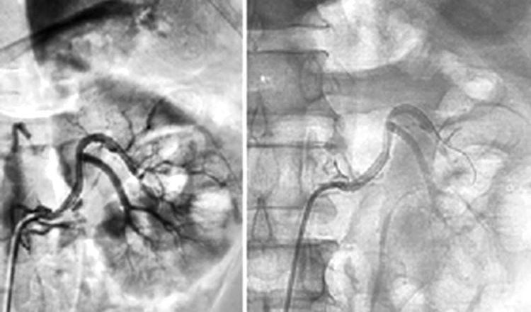
Digital erasure imaging is a combination of conventional angiography by Seldinger technique with computerized image processing techniques. Renal artery angiography (DSA) is an X-ray technique that uses iodinated contrast to brighten blood vessels and show the renal vasculature. This iodine-based dye is completely harmless and will be excreted in the urine.Before the procedure, the machine will take an initial image without the injection as the background image, then the contrast will be injected into the renal artery through a catheter inserted through the skin from the femoral artery. The machine will record a moving image when the contrast material enters the body and process to exclude the background image (digitize and remove the background).
Digital renal angiography with background erasure is indicated in:
● Suspected renal vascular disease: renal stenosis, renal vascular malformation, aneurysm;
Hematuria with suspected renal vascular system;
● Evaluation of blood supply in renal tumor diseases: vascular lipoma;
● Traumatic diseases with suspected renal vascular damage;
● Chronic inflammatory disease with vascular damage;
● Serving interventional radiology;
● Taken to prepare for a kidney transplant.

Người bệnh tiểu ra máu cần được chụp động mạch thận số hoá xoá nền giúp đánh giá tìm nguyên nhân
2. Means and drugs to prepare before taking a renal artery DSA
2.1 Equipment and vehicles ● Digital background eraser angiography (DSA);● Dedicated electric pump;
Film, film printer, image storage system;
● Lead shirt, apron to shield X-rays.
2.2 Medicines to prepare ● Local anesthetic;
● Pre-anesthesia and general anesthetic (if the patient has an indication for general anesthesia);
Anticoagulants;
● Neutralizing anticoagulants;
Water-soluble iodine contrast agent;
● Antiseptic solution for skin and mucous membranes
2.3 Person performing renal artery DSA ● Specialist doctor;
Auxiliary physician;
Electro-optical technician;
● Nursing;
● Doctor, anesthesiologist (if patient cannot cooperate).
Chụp động mạch thận số hoá xoá nền cần được thực hiện bởi bác sĩ chuyên khoa và ekip
● The patient or his/her adoptive family member must write a written consent to the procedure before performing the procedure;
● The patient needs to fast for 6 hours before or drink no more than 50ml of water;
● After entering the intervention room for renal angiography, the patient will lie in the supine position, and the technicians will install a machine to monitor vital signs such as breathing rate, pulse, blood pressure, electrocardiogram, arterial blood gases. In case the patient is uncooperative or aggressive, a sedative will be prescribed;
● Disinfect the skin surface of the groin area with an antiseptic solution and spread a sterile intervention kit.
3. Digital renal angiography with background erasure
3.1 Anesthesia/anesthesia The patient lies supine on the DSA table, the technician places an infusion line, usually 0.9% isotonic saline serum solution is used for infusion. For most cases of renal angiography, the patient only needs local anesthesia.However, in exceptional cases where pre-anesthesia is required, such as: children under 5 years of age, who do not have a sense of cooperation with doctors, or patients who are too excited or scared... then it is necessary to conduct anesthesia. whole body during the procedure.

Người bệnh sẽ được gây tê trước khi tiến hành chụp động mạch thận số hoá xoá nền
● In most cases, it will be inserted through the femoral artery because this is a large artery, easy to insert the catheter.
3.3 Diagnostic renal angiography procedure Sterilize and anesthetize at the puncture site. Insert the needle and insert the catheter into the lumen.
Capture the entire abdominal aorta and bilateral renal arteries by inserting a catheter into the abdominal aorta to the level above the L1 vertebra, then inject contrast media at a rate of 15ml/s, volume pump is 30ml and pump with high pressure 500PSI.
Continue to insert the catheter to the aorta, the level of the L1 vertebrae, then rotate the catheter to the side to hook it to the right renal artery or the left renal artery, then proceed to pump the drug at a rate of 4ml/s, volume pump is 20ml and pump under high pressure 500PSI.
The film focuses carefully on the anterior-posterior renal direction to obtain images of the renal artery, parenchyma, and vein phases. Or you can proceed to shoot 45 degrees to the left.
After the scan, remove the tube from the lumen, apply pressure directly at the needle puncture site for about 15 minutes to stop bleeding, then apply pressure for 8 hours or use an instrument to close the vessel.
Note that during the procedure, it is necessary to monitor the patient's pulse, blood pressure and reaction. After the end, the patient lies on the bed, the leg on the side is performed immobilized, monitoring the blood vessels in the instep of the foot on the side that the catheter is inserted, monitoring for bleeding and hematoma at the needle puncture site. , control systemic signs: heart, pulse, blood pressure.
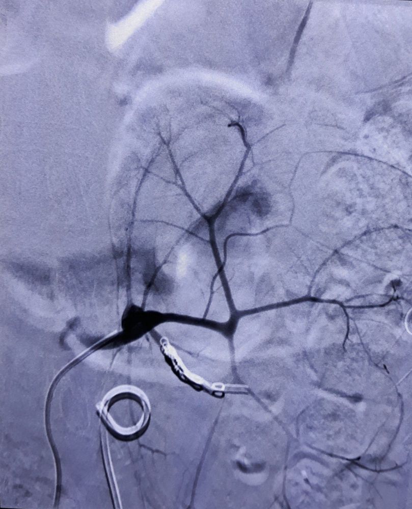
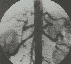
Hình ảnh nút động mạch thận trên công nghệ chụp mạch số hóa xóa nền (DSA) trên bệnh nhân tiểu ra máu
4. Complications and how to handle them
4.1 Complications during the procedure ● Tear the artery wall causing bleeding or dissection of the artery wall at the catheter insertion site. Treatment is by stopping the procedure immediately, applying local compression and monitoring closely. Continuation of the scan by puncture of the contralateral artery;● Break the catheter, guide and save the broken piece in the lumen: use special tools to remove it through endovascular or surgical intervention;
● Allergy to contrast: early diagnosis and treatment of contrast agent complications.
4.2 Complications after the procedure ● Bleeding at the catheter insertion site: Apply pressure at the site with correct technique and remain motionless until the bleeding stops;
● Infection: Using antibiotics to treat infections because this is an invasive method, there is still a risk of infection;
● If bulging or arteriovenous septal defect occurs, catheter rupture or lead rupture (although rare) requires endovascular or surgical management.
Digitalized limb angiography to remove the background gives the best results when the medical facility is equipped with modern and advanced equipment. Therefore, you should visit reputable and high-quality medical facilities.
Vinmec International General Hospital is a high-quality medical facility in Vietnam with a team of highly qualified medical professionals, well-trained, domestic and foreign, and experienced. A system of modern and advanced medical equipment, possessing many of the best machines in the world, helps to detect many difficult and dangerous diseases in a short time, supporting the diagnosis and treatment of effective doctors. most fruitful. The hospital space is designed according to 5-star hotel standards, giving patients comfort, friendliness and peace of mind.
Master. Dr. Le Hong Chien has many years of experience working in the field of diagnostic imaging and interventional radiology (endovascular intervention and extravascular intervention). Before being a Doctor of Diagnostic Imaging - Intervention at Vinmec Times City International Hospital, Dr. Chien was a Doctor at the Department of Diagnostic Imaging, Hospital 19.8 - Ministry of Public Security and Hong Ngoc General Hospital.
To register for an examination at Vinmec International General Hospital, you can contact the nationwide Vinmec Health System Hotline, or register online HERE.


