This is an automatically translated article.
The article was professionally consulted by Specialist Doctor I Tran Cong Trinh - Radiologist - Radiology Department - Vinmec Central Park International General Hospital.
Abdominal aortic angiography allows to clearly see the structure of the abdominal aorta, is indicated in cases of abdominal trauma, suspected abdominal aortic stenosis, ...
1. What is abdominal aortic angiography?
Abdominal aortic angiography by digitizing background erasure machine using iodinated contrast is a technique that allows to clearly visualize the abdominal aorta structure, the segment from below the diaphragm to the bifurcation of the main iliac arteries. The scan results show that the arteries include: visceral artery, renal artery, superior mesenteric artery, lower colon, hypogastric artery, bilateral main iliac artery, double intercostal arteries originating from abdominal aorta.2. Who is indicated for abdominal aorta?
Abdominal aortic angiography is indicated for the following cases:Congenital disease and pathologies related to the abdominal aorta such as: abdominal aortic aneurysm, abdominal artery stenosis, arteriovenous fistula, mass angiomas, underdeveloped vessels, ... Accidental trauma to the abdomen and/or lungs, suspected vascular injury. Tumor in the abdomen suspected of compression, invasion causing damage to the vessels. Serving organ transplant surgery or radiological intervention.
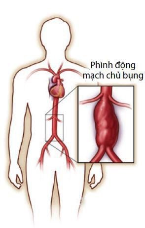
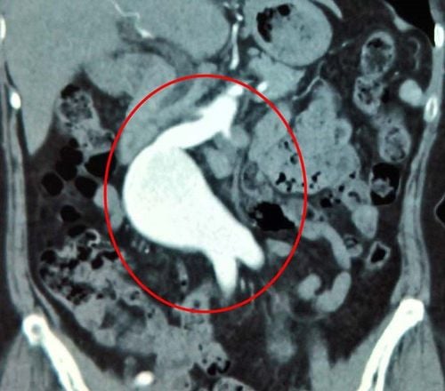
Bệnh nhân phình động mạch chủ bụng được chỉ định thực hiện
3. How is an abdominal aortic angiogram using a background eraser digitizer done?
Abdominal aortic angiography by digitizing background eraser is performed with the following steps:Step 1: Perform anesthesia by placing the patient supine on the table, setting up an intravenous line, and anesthetizing at place. In cases where the patient is agitated, scared, unable to cooperate like a child under 5 years old, a pre-anesthetic drug can be injected to induce general anesthesia before the procedure. Step 2: Select the location and technique to insert the catheter into the artery. Usually, the catheter is inserted from the femoral artery and the technique used is Seldinger. If access from the femoral artery is not possible, substitute access from the axillary, brachial, or radial artery. Disinfect, anesthetize the place where the needle needs to be inserted to put the tube into the artery.

Kết quả hình ảnh chụp động mạch chủ bụng
Abdominal aortic angiography is a modern imaging technique that helps to detect lesions in the abdominal aorta, thereby providing appropriate treatment.
Digital scan of the abdominal aorta is a modern imaging technique that helps doctors detect the location and extent of the disease. However, for accurate results, patients should choose reputable addresses with modern DSA scanners, and at the same time, it must be performed by qualified technicians and doctors.
Vinmec International General Hospital has applied digital imaging technology to erase the background in examination and diagnosis of many diseases. The process of digitizing and erasing the background at Vinmec is carried out methodically and according to the standards of the process by a team of highly skilled doctors and modern machinery, thus giving accurate results, making a significant contribution. in determining the disease and the stage of the disease for timely treatment.
Before taking a job at Vinmec Central Park International General Hospital, the position of Doctor of Radiology from September 2017, Doctor Tran Cong Trinh worked at Gia Dinh People's Hospital since 2007. -2017. In his role, Dr. Tran Cong Trinh has participated in guiding the teaching of students, residents, specialists and new doctors entering the department
For examination and treatment at International General Hospital Vinmec, please come directly to Vinmec Health System or register online HERE.


