This is an automatically translated article.
The article was professionally consulted by Specialist Doctor I Tran Cong Trinh - Radiologist - Radiology Department - Vinmec Central Park International General Hospital.
Pelvic angiography with digital background eraser is a technique that allows the detection of pelvic trauma or pelvic tumor. This is a modern imaging method used in many medical facilities and hospitals.
1. Digital iliac angiography with background erasure is what technique?
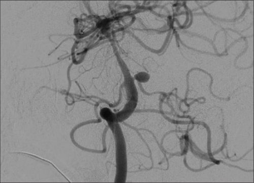
Pelvic angiography by digitizing background ablation machine using iodinated contrast is a technique that allows visualization of the abdominal aorta and iliac arteries including the main iliac, internal and lateral iliac arteries.This technique is indicated for pelvic pathology and trauma (including abdominal aorta, iliac root, internal and external iliac), tumors in the pelvic region. In addition, this technique also serves electro-optical intervention.

Chấn thương vùng chậu gây tổn thương mạch có thể được phát hiện bằng chụp số hóa xóa nền động mạch chậu
2. How is background digitized iliac angiography performed?
Digital iliac angiography with background erasure is performed including the following steps:Step 1: Anesthetize the patient by placing the patient supine on the examination table, placing an intravenous line, and administering local anesthesia. In cases of excitement, fear, inability to cooperate, pre-anesthesia can be injected. Step 2: Sterilize and anesthetize the femoral artery, insert the needle and use the Seldinger technique to insert the catheter into the artery. If access from the femoral artery is not possible, the axillary, brachial, or radial artery can be substituted. Step 3: After inserting the catheter, perform injection and iliac angiography. Depending on the segment to be taken, the drug will be injected at different speeds and amounts. With the terminal segment of the abdominal aorta and the bilateral external iliac arteries, insert the 5F (Pigtail) tube to the level of the superior border of L4, then use a high-pressure iodinated contrast injection pump (800 PSI). , speed 1015ml/s. With the main iliac artery segment, the internal or external iliac on each side, insert the 5F Cobra tube to the main iliac artery on each side, then use an iodinated contrast injection pump with an amount of about 15ml at 500 PSI pressure, at 5ml/s . Step 4: Record and shoot continuously, focusing on shooting in the subframe from the level of L3 to below the pubic bone on both sides. For a clear and accurate result, the iliac and femoral arteries can be taken at 25 - 300 angles. Step 5: After the iliac angiogram is satisfactory, proceed to withdraw the catheter and tube into the lumen, stop bleeding by directly pressing on the needle puncture site for 15 minutes, then use a closing device. intravascular or compression bandage for 6 hours. The resulting image must clearly show the structure, the aorta-iliac system including the main iliac arteries, the internal iliac and the external iliac on both sides, and at the same time allow to detect lesions (if any).
3. Management of complications during and after iliac angiography
During and after iliac angiography, there may be some complications that need to be handled appropriately and promptly:Bleeding: During the procedure, at the needle puncture site may appear a condition. bleeding from an artery laceration or dissection. To stop bleeding, it is necessary to stop the procedure and apply pressure by hand, then bandage to monitor, redirect to the opposite side. After the scan, there may be bleeding or hematoma at the catheter site, which should be treated by applying pressure and continuing to lie motionless to monitor until the bleeding stops. Fractures, breaks, intravascular instruments: Endovascular intervention or surgery using special instruments to remove. Arterial occlusion: During lower extremity angiography, blockage of an artery due to a blood clot or plaque detachment can occur. At this point, the specialist can treat it with surgical intervention to remove the clot or inject fibrinolytic drugs. Angioplasty or catheterization: Endovascular intervention or surgical treatment. Infection: Treat with antibiotics.

Tắc động mạch có thể xảy ra sau khi chụp động mạch chậu
Digitization of the iliac artery background is a modern imaging technique that helps doctors detect the location and extent of damage to the disease. However, for accurate results, patients should choose reputable addresses with modern DSA scanners, and at the same time, it must be performed by qualified technicians and doctors.
Vinmec International General Hospital has applied digital imaging technology to erase the background in examination and diagnosis of many diseases. The process of digitizing and erasing the background at Vinmec is carried out methodically and according to the standards of the process by a team of highly skilled doctors and modern machinery, thus giving accurate results, making a significant contribution. in determining the disease and the stage of the disease for timely treatment.
Before taking a job at Vinmec Central Park International General Hospital, the position of Doctor of Radiology from September 2017, Doctor Tran Cong Trinh worked at Gia Dinh People's Hospital since 2007. -2017. In his role, Dr. Tran Cong Trinh has participated in guiding the teaching of students, residents, specialists and new doctors entering the department
For examination and treatment at International General Hospital Vinmec, please come directly to Vinmec Health System or register online HERE.


