This is an automatically translated article.
The article was professionally consulted by Specialist Doctor I Tran Cong Trinh - Radiologist - Radiology Department - Vinmec Central Park International General Hospital.Renal vascular malformation is one of the rare vascular malformations that cause hematuria. Because of the abnormality in blood vessels, parenchymal structure and kidney function are also affected. Accordingly, the digital scan to erase the background and node of the renal vessel malformation is a radical intervention, restoring the normal vascular structure of the kidney and preserving kidney function in the long run.
1. What is a renal vascular malformation?
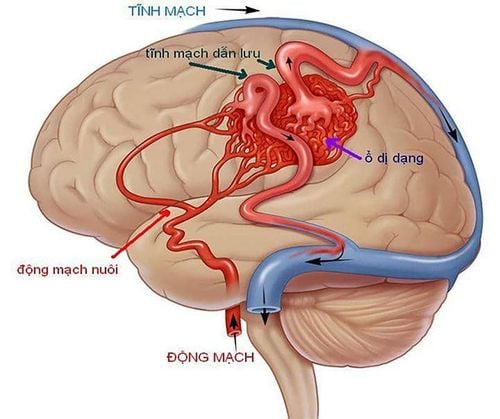
When there is an abnormal connection between the arteries and veins outside the capillary level, it is called an arteriovenous malformation. This is the result of vascular development during the incomplete embryonic stage, usually occurring between the 10th week. Besides, renal vascular malformation can also be the result of trauma to the renal parenchyma. such as kidney injury or kidney biopsy. Renal vascular malformations occur more frequently in women than in men (3:1) and usually involve the right kidney.The most common clinical presentation of renal vascular malformation (75% of cases) is microscopic hematuria or gross hematuria. Hematuria occurs as a result of dysplastic blood vessel rupture in the urine collection system and can become a life-threatening problem in many cases of major blood loss. The severity of hematuria is not related to the size of the lesion; Even small renal vessel malformations can lead to severe blood loss if located near the pelvic artery system. If the rate of blood loss is slow, blood clots can form, obstructing the urinary tract and causing flank pain. In addition, some patients also show signs of hypertension, congestive heart failure or early renal failure, especially in cases of congenital renal vascular malformation. Whatever the cause, such complex lesions need to be confirmed and definitive treatment instituted as soon as possible to preserve renal function. In which, digital scan to erase the background and node malformation of the kidney is one of the preferred methods to choose.

Tiểu ra máu là triệu chứng phổ biến của bệnh
2. What is a digitized scan of the background and node of renal vascular malformation?
Once the patient presents with hematuria and the diagnosis of renal vascular malformation has been established, an urgent treatment plan needs to be in place, especially when hematuria is life-threatening. Surgical options include the highly invasive total or partial nephrectomy, the risk of complete loss of renal function, and the risk of herniation of the lateral abdominal wall. Therefore, digital angiography and renal vascular malformation as a percutaneous interventional procedure have been developed to manage such complex lesions.Renal artery malformation node procedure is usually performed under local anesthesia. The goal is to permanently block with material that obstructs the small channels between the arteries and veins that form the anastomosis in renal vascular malformations. The entire manipulation process is in the lumen of the artery. The outcome of the procedure also depends on the skill of the operator and the type of blockage material used. Commonly used materials are gel foam, pure alcohol, polyvinyl alcohol, metal coils or bio-liquid adhesives. Sometimes there is a combination of two materials at the same time, depending on the anatomical features of the deformity.

Khi thực hiện thủ thuật, bệnh nhân chỉ cần gây tê tại chỗ
3. What is the process of digitizing the background and node of renal vascular malformation?
Patients and relatives need to be thoroughly explained about the procedure to be prepared, as well as the risks involved, in order to ask for cooperation. Next, the patient needs to fast, change into appropriate clothes and arrange to lie on his back on the intervention table, place an intravenous line with 0.9% physiological saline solution.Means of monitoring breathing, pulse, blood pressure, electrocardiogram, SpO2 are mounted on the patient and the indicators are displayed on the screen for easy observation. After cleaning the inguinal and genital areas, the doctor will cover the area with a sterile tissue to prepare for intervention. In case the patient is overstimulated and poorly cooperated, it is necessary to consider appointing a sedative to facilitate the process.
At the intervention site, the doctor will administer local anesthesia from the femoral artery. When the anesthetic takes effect, the doctor will insert a needle and insert the catheter into the artery, leading to the location of the abnormal vertebral artery. Here, the doctor will conduct an assessment of the injury by taking an abdominal aorta from the base of the renal artery with a pigtail catheter. Next, the renal artery on the affected side was also selectively imaged with a Cobra catheter.
Once the lesion is located, the doctor will approach by inserting a catheter into the root of the ipsilateral renal artery. Then, the catheter and micro-conductor will be inserted into the pedicle of the blood vessel that feeds the malformation. A volume of contrast agent was injected for angiography to locate the tip of the microcatheter that was already in the fovea of the arteriovenous malformation. Under the background removal digitizer screen, the doctor will remove the micro-conductor and replace it with a rigid micro-conductor to begin treatment with the plug material. Depending on the type and approach to the deformed drive, select the appropriate plug material. Metal spirals or bio-glue are two commonly used materials. However, the choice of material depends on the availability and experience of the surgeon.
To end the procedure, your doctor will need an angiogram. Determining whether the results are satisfactory or not is based on the DSA images taken at the location of the bottleneck, adjacent branches and downstream. The deformed stopper must be in good working order, with no backflow and no displacement. At the same time, the doctor will also check that the pulse before, during and after the blockage is still circulating normally, there are no signs of thrombosis or dissection of the vessel wall. When the above conditions are ensured, the interventional tools on both sides will be withdrawn. The puncture artery site will be directly pressed by hand for about 15 minutes to stop bleeding and then bandaged for 8 hours. During this time, the patient is limited to bed movement.
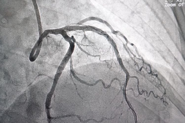
Trên hình ảnh của DSA, bác sĩ sẽ đánh giá được kết quả của thủ thuật
4. What complications can be encountered when digitizing the background and node malformation of the kidney?
Similar to any intracranial interventional procedure, digital angiography and renal vascular malformation can also carry certain risks, including:Arterial tear causing intra-abdominal bleeding Dissection Arterial wall displacement of the embolus causing renal or mesenteric infarction Hematoma or bleeding at the puncture site Infection at the puncture site Complications related to anesthesia, anesthesia, or contrast media use.
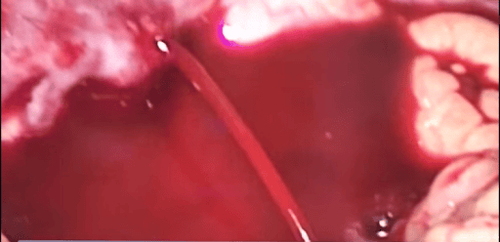
Chảy máu ổ bụng có thể xảy ra khi chụp số hóa xóa nền và nút dị dạng mạch thận
Doctor Tran Thi Mai Huong has 25 years of experience in examination and treatment in the field of Obstetrics and Gynecology, lower tract surgery, laparoscopic surgery. Has held the position of deputy head of the School of Obstetrics and Gynecology, deputy head of the delivery department - Hai Phong Obstetrics and Gynecology Hospital.
For examination and treatment at Vinmec International General Hospital, please come directly to Vinmec Health System or register online HERE.


