This is an automatically translated article.
The article was professionally consulted by Master, Doctor Pham Manh Chung - Department of Diagnostic Imaging - Vinmec Ha Long International HospitalBreast cancer screening is a measure to help detect the disease early, increase treatment efficiency, and reduce costs and time. In disease screening methods, imaging plays an important role in diagnosing and detecting abnormalities in the mammary glands, especially breast cancer.
1. Breast Cancer Overview
Breast cancer is the most common cancer and the leading cause of death in women. The cause of breast cancer is still not clear. However, many studies suggest that BRCA1 or BRCA2 gene mutations, environmental pollution, radiation, chemicals in food, smoking habits,... increase the risk of breast cancer.
Breast cancer patients can be discovered by chance through routine examination or when clinical signs appear such as: There is a firm mass in the breast, no pain, no movement. Late signs of the disease include breast skin changes, breast bleeding, pectoral lymphadenopathy, and axillary fossa. Warning symptoms of metastatic disease include pleural effusion, supraclavicular lymphadenopathy, anemia, bone pain, fracture, paralysis, limb weakness, coma due to brain metastases,...

Tầm soát ung thư vú
2. Why is breast cancer screening necessary?
Today, with the development of modern techniques, screening measures and early detection of breast cancer help improve the cure rate for breast cancer patients. Breast cancer patients when detected at stage 1 and actively treated have an 80-90% chance of survival for more than 5 years. Therefore, screening and screening for breast cancer is extremely important in detecting the disease early and improving the effectiveness of disease prevention and treatment.
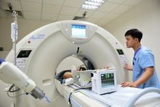
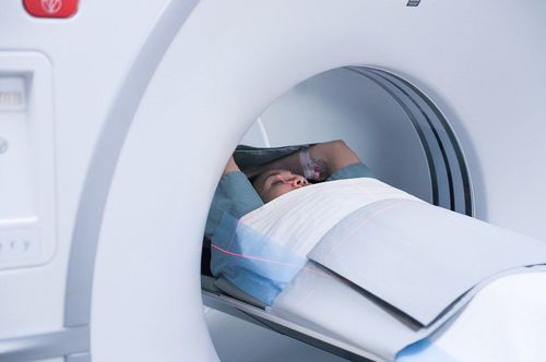
Commonly used methods in breast cancer screening programs are clinical breast exams (done annually for women over 40) or breast self-exams at home. In addition, women are also advised to perform imaging in breast cancer pathology. Commonly used imaging methods include mammography (mammography), ultrasound, computed tomography (CT scan) and magnetic resonance imaging (MRI).
3. Imaging methods for breast cancer

Dấu hiệu cảnh báo sớm bị ung thư vú
3.1 Mammography (mammography)
This is a special mammogram, which plays an important role in breast cancer screening. This is a procedure that uses low-intensity X-rays to shine on the mammary gland tissue to capture images of the mammary gland. Through images obtained from mammograms, doctors can detect abnormalities and tumors at an early stage even when the patient has not seen or felt them.
Mammography technique has outstanding advantages such as fast implementation time, non-invasive, accurate results, reasonable cost,... Many studies show that Mammography helps to reduce the mortality rate of cancer. breast cancer about 30%. Therefore, oncologists prefer to specify this imaging technique. Women over the age of 40 should have mammograms 1-2 times a year.
Regarding the meaning of screening and diagnosis, mammograms bring specific values such as:
Detecting abnormal lesions in the breast, axillary fossa on both sides; Detect signs of microcalcification that cannot be detected by ultrasound - one of the malignant signs of mammary disease; Breast cancer risk screening; Early diagnosis of breast cancer, including tumors that are not palpable on physical examination, inconspicuous lesions, intraluminal lesions, and very small calcified lesions with a sensitivity of over 90% ; Monitor known lesions, detect recurrence or new lesions in cases of breast tumor surgery; Guide to breast biopsy; Follow-up breast cancer treatment. 3.2 Breast ultrasound
Breast ultrasound is a method of imaging breast cancer by building and reproducing images of the internal structure of the breast and body. Breast ultrasound also supports needle guidance in invasive procedures such as: Aspiration cyst, aspiration biopsy, guided biopsy needle with core,... This method is being applied quite popularly because of its low cost. low cost, easy to perform, painless and harmless to the patient. Ultrasound diagnosis of breast disease has high accuracy, can diagnose small lesions less than 5mm in diameter, valuable in early breast cancer detection.
The advantage of ultrasound over mammography is that it avoids the patient's exposure to X-rays. It can diagnose pregnant women, X-ray-sensitive patients, children in puberty, and patients. have enlarged, thickened mammary glands (mammography does not clearly identify lesions). In addition, ultrasound also helps patients feel more comfortable because there is no pressure on the breast during mammography.
With the development of science and technology, ultrasound machines are increasingly integrated with many modern technologies. The introduction and development of ultrasound elastography increases the diagnostic value of ultrasound in the diagnosis of breast cancer.
3.3 Mammography MRI
Magnetic resonance imaging is also a modern imaging technique to help detect cancer and some other abnormalities in the mammary gland with high accuracy. The image will be displayed on the computer for the doctor to make an accurate conclusion.
4. Who should have breast cancer imaging?
Women between the ages of 20 and 30: Should start screening for breast cancer by self-examination, specialist examination every 3 years and up to age 40 should have a specialist examination once a year; Women 40 years of age and older: Breast cancer should be screened by mammography or mammography once a year; Women at high risk for breast cancer should have an ultrasound, mammogram, and magnetic resonance imaging once a year. High risk factors include: Family history of breast cancer, early menarche (before age 12), late menopause (after age 55), regular use of oral contraceptives or estrogen replacement therapy. , have no children or give birth to their first child after the age of 30, have a diet high in animal fat, have a habit of smoking, drinking alcohol,...; Mammogram and breast ultrasound immediately when there are abnormal breast symptoms such as palpating solid mass, skin or nipple retraction, nipple discharge, breast skin color change,... Diagnostic methods The above image diagnosis helps to screen for breast diseases, especially early breast cancer, to help guide doctors in timely diagnosis and treatment, and to improve treatment efficiency for patients.
Vinmec International General Hospital is currently implementing 4 packages of breast cancer screening and screening for many subjects. The examination packages include: Basic package, standard package, advanced package and intensive package, hierarchical according to each group of subjects with increasing risk of breast cancer. With a team of experienced, highly qualified specialists and a system of modern medical equipment, patients will be diagnosed with high accuracy about their health problems and receive advice. best in the prevention and treatment of diseases.
Please dial HOTLINE for more information or register for an appointment HERE. Download MyVinmec app to make appointments faster and to manage your bookings easily.