
This is an automatically translated article.
The article was consulted with Master, Doctor Tran Hong Nhat - Interventional Cardiologist - Cardiovascular Center - Vinmec Central Park International General Hospital.Coronary artery failure leading to myocardial ischemia can be chronic (chronic coronary syndrome) or acute (acute coronary syndrome). Diagnosis of myocardial ischemia is based on clinical symptoms and biochemical blood tests as well as cardiac imaging.
1. Myocardial ischemia
Myocardial ischemia occurs when there is insufficient blood flow to an area of the heart due to narrowing of the coronary arteries that supply that area. Although coronary artery stenosis can be caused by vasospasm, it is most often caused by the accumulation of plaque or by a blood clot that narrows or blocks the lumen of the blood vessel. Most people with early-stage ischemic heart disease (less than 50% stenosis) usually have no symptoms. However, as atherosclerosis progresses, especially if left untreated, symptoms can occur.Diagnosis of myocardial ischemia can be done in several ways. If the patient has had a previous myocardial infarction or has undergone coronary revascularization (by PCI or CABG), there is myocardial ischemia. Furthermore, the presence of typical angina suggests a clinical diagnosis, but most often requires confirmation by additional diagnostic tests, such as echocardiography, coronary angiography, and angiography. Coronary CT, cardiac MRI.
2. Clinical symptoms of myocardial ischemia
Symptoms of ischemic heart disease often occur with exercise, when the demand for oxygen carried by the blood increases. The discomfort in the chest when the heart muscle is deprived of oxygen is called angina. This is a clinical syndrome characterized by a feeling of heaviness in the chest, lasting 5-10 minutes, which may radiate to the jaw, shoulder, back, or arms, usually aggravated by exertion or stress and relieved by rest, take nitroglycerin.Okay. Patients with normal coronary arteries may also have angina associated with coronary vasospasm or endothelial dysfunction.
Angina or symptoms equivalent to angina (eg, dyspnea on exertion) classified based on the description of the level of activity causing the symptoms according to the Canadian Heart Association (CCS) ):
Type I: Angina occurs with intense, rapid or prolonged exertion, but does not occur with usual physical activity. Type II: Angina that slightly limits normal activities, such as angina that occurs when walking or climbing stairs quickly, walking uphill, walking or climbing stairs after a meal; in cold weather or in the wind; emotionally stressed; only in the first few hours after waking up; walk more than two blocks on level ground and climb more than one normal flight of stairs at a normal pace and under normal conditions. Type III: Angina markedly limiting normal physical activity, such as angina that occurs when walking a block or two on level ground, climbing a flight of stairs in normal conditions and at a normal pace, housework, gardening, vacuuming... Class IV: Angina that renders the inability to perform any physical activity without discomfort; Angina syndrome may occur at rest.

Cơn đau thắt ngực là một trong những triệu chứng của thiếu máu cục bộ cơ tim.
3. Subclinical diagnosis of myocardial ischemia
Blood biochemical tests Metabolic disorders are common in patients with myocardial ischemia, so it is necessary to check fasting blood glucose; blood lipids (total cholesterol, LDL-cholesterol, HDL-cholesterol, triglycerides); liver enzymes; blood creatinine; estimated glomerular filtration rate (eGFR) and hs-CRP. HS-CRP.Biomarkers of cardiac enzymes (Troponin, CKMB) increased indicating myocardial damage in patients with ischemic heart disease, helping to differentiate acute coronary syndromes from other causes
Electro ECG Several abnormalities on the electrocardiogram contribute to the diagnosis of myocardial ischemia, such as: Q waves of an old myocardial infarction, abnormal ST-segment elevation or depression, or new left bundle branch block. presently.
Echocardiography Echocardiography is a convenient and useful tool for patients with suspected myocardial ischemia. Echocardiography helps to identify motor abnormalities in suspected coronary artery disease, evaluate left ventricular ejection fraction for risk stratification, and assess left ventricular diastolic function.
Signs of regional dyskinesia, such as hypoactivity, inactivity, or paradoxical movement (dyskinesia), combined with signs of ventricular septal wall not thickening during systole, suggest a diagnosis of myocardial ischemia. Local. In particular, when dyskinesia and reduced ejection fraction occur during exercise testing with a wheelchair, bicycle, or medication.
Stress test Is a non-invasive means of diagnostic and prognostic value.
An exercise electrocardiogram (ECG) on a bicycle or roller mat is a common and safe test. The most important diabetic changes are ST-segment depression and ST-segment elevation. Stress ECG is positive when ST depression is horizontal or depressive > 1mm lasting 0.06 - 0.08 seconds after the end of the QRS during or after exercise.
Stress echocardiography (SAT) and exercise stress cardiography (roller mat, bicycle) or drug (dobutamine, adenosine, dipyridamole) have higher sensitivity and specificity than exercise diabetes. Physical exertion (6-12 minutes) is preferred over medication. However, in patients who are unable to exercise, either stress SAT or pharmacological stress radiography is required. Stress SAT signs suggestive of myocardial ischemia include: Decreased wall motility > 1 region, decreased wall thickening > 1 region, compensatory hyperactivity in non-ischemic myocardium.
Computed tomography (MSCT) The MSCT scan helps to calculate the coronary calcification score and visualize the coronary arteries if contrast is used. The coronary calcification score has high sensitivity (90%), but low specificity in coronary artery disease (50%). Currently, coronary MSCT angiography is not routinely indicated for the screening of coronary heart disease in individuals at low risk of myocardial ischemia (estimated 10-year risk < 10%). Patients with confirmed coronary artery disease, exercise DM is better than MSCT to decide before coronary angiography. An MSCT coronary angiogram is indicated when a patient is at moderate risk for coronary artery disease and is symptomatic, especially when exercise testing is inconclusive. Coronary MSCT has a high negative predictive value, so it can be done in cases where coronary artery disease needs to be ruled out.
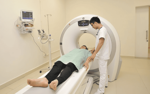
Chụp MSCT là một trong những phương pháp chẩn đoán thiếu máu cục bộ cơ tim.
Coronary angiography Coronary angiography is considered the gold standard in the diagnosis of myocardial ischemia. This is the most accurate, invasive exploratory method to confirm the diagnosis of coronary artery occlusion due to atherosclerosis, coronary vasospasm, Kawasaki disease, coronary artery dissection, and coronary artery disease due to radiotherapy
In a nutshell, myocardial ischemia is recurrent chest pain or discomfort that occurs when part of the heart doesn't get enough blood and oxygen. It is usually caused by coronary atherosclerosis, which narrows the lumen of the vessel, eventually obstructing the flow. Diagnosis of myocardial ischemia is based on angina symptoms, cardiac markers, electrocardiogram, and imaging tests such as echocardiography. When silent myocardial ischemia or coronary artery disease is suspected, cardiovascular imaging such as coronary computed tomography, cardiac MRI, stress echocardiography... plays an important role.
Currently, Cardiovascular Center - Vinmec International General Hospital is one of the leading centers in the country for examination, diagnosis, screening and treatment of cardiovascular diseases. Vinmec not only has the convergence of a team of experienced and reputable leading experts in the field of surgical treatment, internal medicine, interventional cardiac catheterization, but also has a system of modern equipment, on par with The most prestigious hospitals in the world such as: MRI 3 Tesla (Siemens), CT 640 (Toshiba), high-end endoscopy equipment EVIS EXERA III (Olympus Japan), high anesthesia system Avace level, Hybrid operating room according to international standards... Especially, with the space designed according to 5-star hotel standards, Vinmec ensures to bring patients the most comfort, friendliness and peace of mind. .
Please dial HOTLINE for more information or register for an appointment HERE. Download MyVinmec app to make appointments faster and to manage your bookings easily.